Imaging Findings of Breast Metastasis from Squamous Cell Carcinoma of the Cervix: A Case Report
- Affiliations
-
- 1Department of Radiology, Soonchunhyang University Seoul Hospital, Seoul, Korea. ywchang@schmc.ac.kr
- 2Department of Pathology, Soonchunhyang University Seoul Hospital, Seoul, Korea.
- KMID: 2442473
- DOI: http://doi.org/10.3348/jksr.2019.80.1.135
Abstract
- Metastasis from extramammary malignancy to the breast is rare, and metastasis of cervical cancer to the breast is quite uncommon. We report atypical sonographic findings of a rapid growing, single, and circumscribed mass with complex cystic and solid echo pattern in a 50-year-old female. The mass confirmed a metastasis from cervical cancer. It is rare, but the possibility of breast metastasis should be considered when a rapidly growing breast mass is located in between the parenchyma and subcutaneous fat layer.
MeSH Terms
Figure
-
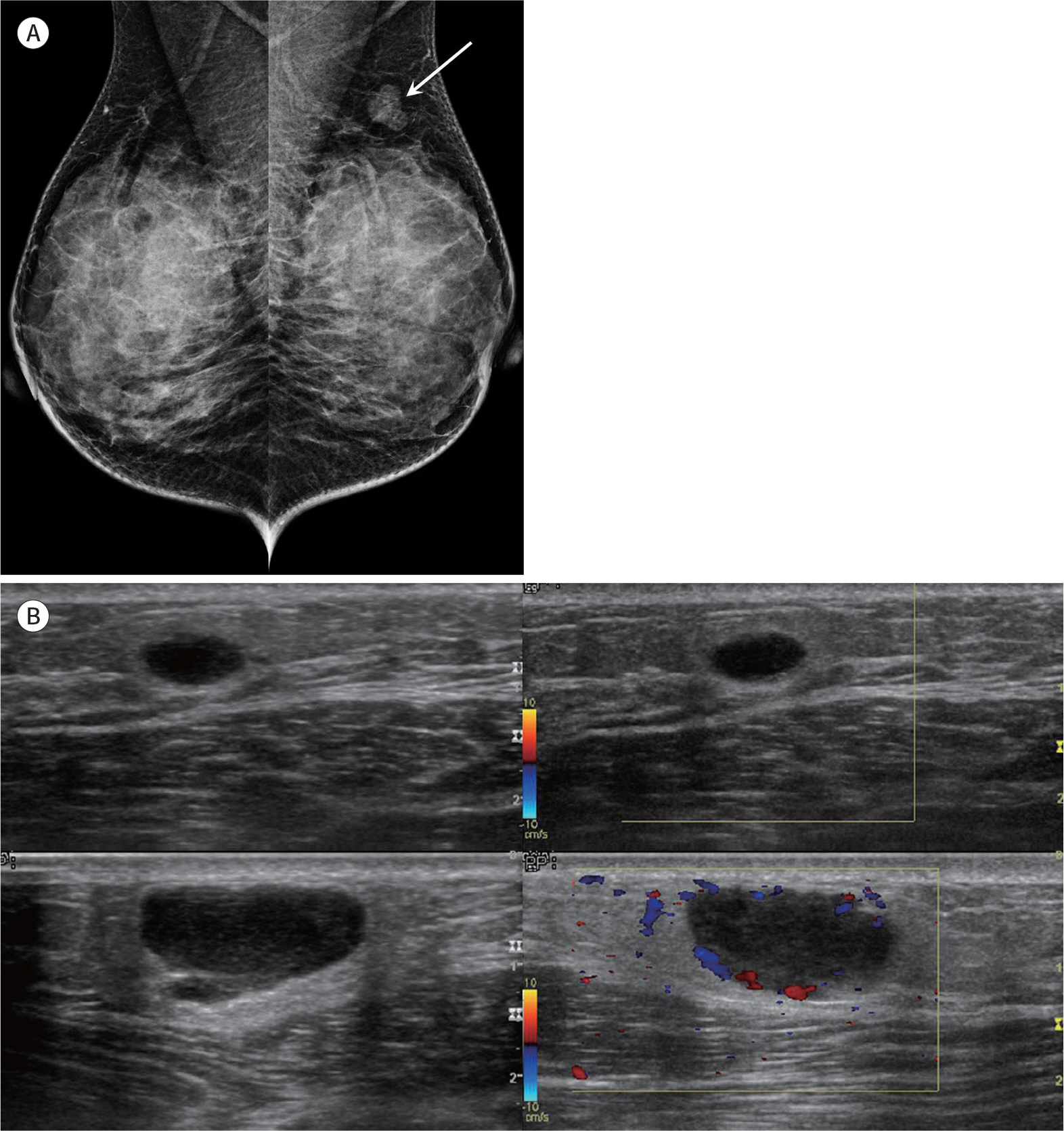
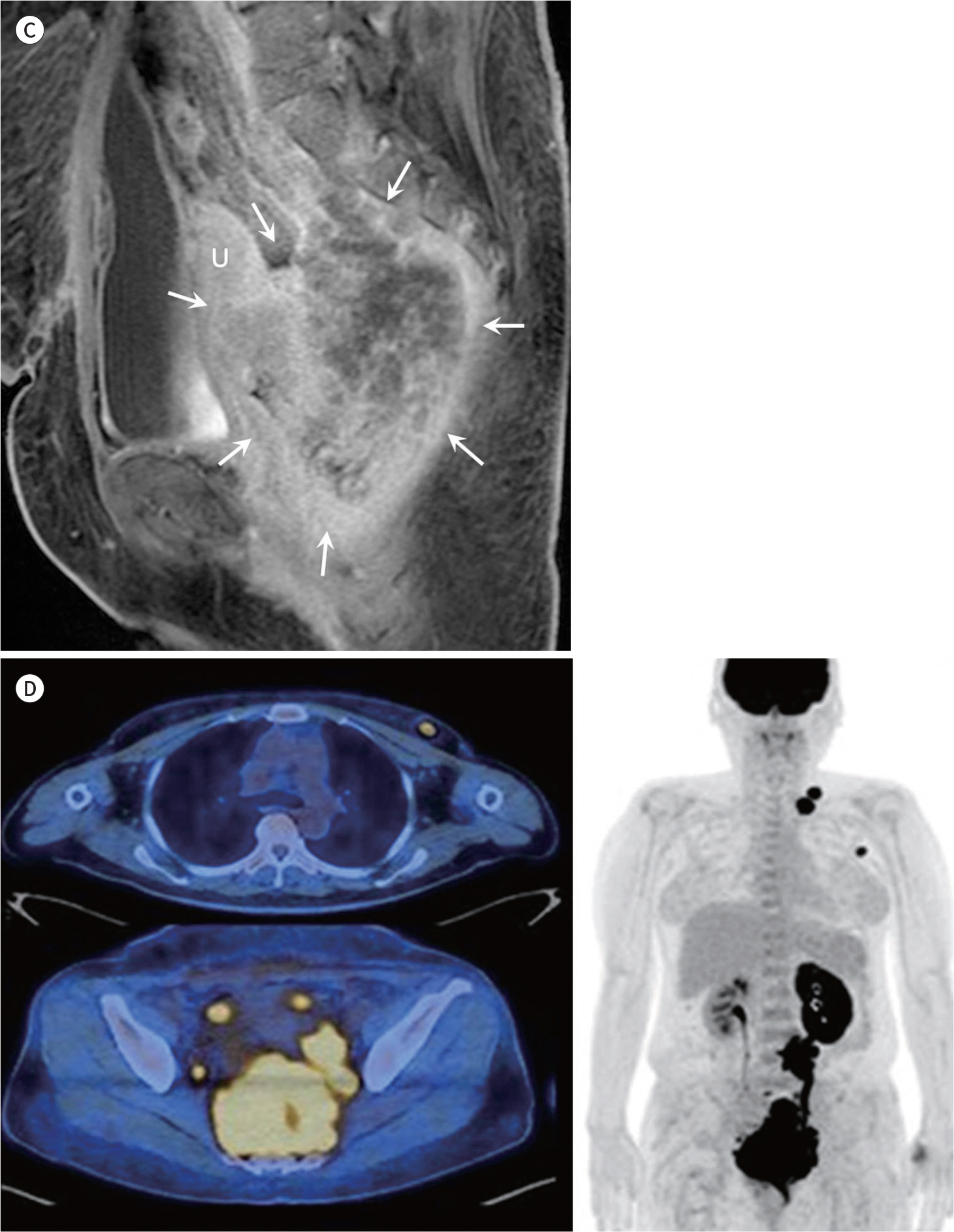
Fig. 1. A 50-year-old woman with breast metastasis from squamous cell carcinoma of cervix. A. Mammogram shows an oval, circumscribed, and equal-density mass in the upper portion of the left breast (arrow). B. An oval, circumscribed, hypoechoic mass is noted between the subcutaneous fat layer and parenchyma at the left breast on ultrasonography and no vascularity on color Doppler image (right upper and leftupper panel, 0.97 × 0.49 × 1.76 cm). After 3 weeks, the mass is a larger, oval, circumscribed, complex cystic and solid mass with increased peripheral vas-cularity (left lower and right lower panel, 2.14 × 1.04 × 2.41 cm). A 50-year-old woman with breast metastasis from squamous cell carcinoma of cervix. C. Enhanced pelvic MRI shows heterogeneous enhancing mass (arrows) at the uterine cervix, invading rectum, upper vagina, and left ureter. D. PET-CT scan shows a hypermetabolic mass at the uterine cervix, invading rectum, upper vagina, and left ureter. Peritoneal seeding nodules and metastatic lymphadenopathy at the left paraaortic and subclavian area are apparently disseminated disease. The breast mass shows hypermetabolism on the PET-CT scan. U = uterus
Reference
-
References
1. Mangla A, Agarwal N, Saei Hamedani F, Liu J, Gupta S, Mullane MR. Metastasis of cervical cancer to breast: a case report and review of literature. Gynecol Oncol Rep. 2017; 21:48–52.
Article2. Bartella L, Kaye J, Perry NM, Malhotra A, Evans D, Ryan D, et al. Metastases to the breast revisited: radiological-histopathological correlation.Clin Radiol. 2003; 58:524–531.3. Gupta S, Gupta MK, Gupta R, Mishra RS. Breast metastasis of cervical carcinoma diagnosed by fine needle aspiration cytology. A case report. Acta Cytol. 1998; 42:959–962.4. Mun SH, Ko EY, Han BK, Shin JH, Kim SJ, Cho EY. Breast metastases from extramammary malignancies: typical and atypical ultrasound features.Korean J Radiol. 2014; 15:20–28.5. Vizcaíno I, Torregrosa A, Higueras V, Morote V, Cremades A, Torres V, et al. Metastasis to the breast from extramammary malignancies: a report of four cases and a review of literature.Eur Radiol. 2001; 11:1659–1665.6. Bohman LG, Bassett LW, Gold RH, Voet R. Breast metastases from extramammary malignancies. Radiology. 1982; 144:309–312.
Article7. Lee SH, Park JM, Kook SH, Han BK, Moon WK. Metastatic tumors to the breast: mammographic and ultrasonographic findings.J Ultrasound Med. 2000; 19:257–262.8. Vergier B, Trojani M, de Mascarel I, Coindre JM, Le Treut A. Metastases to the breast: differential diagnosis from primary breast carcinoma. J Surg Oncol. 1991; 48:112–116.
Article9. McCrea ES, Johnston C, Haney PJ. Metastases to the breast. AJR Am J Roentgenol. 1983; 141:685–690.
Article
- Full Text Links
-
- Actions
-
Cited
- CITED
-
- Close
- Share
-
- Similar articles
-
- A Case of Duodenal Metastasis from Squamous Cell Carcinoma of the Uterine Cervix
- Squamous Cell Carcinoma of the Cervix with Intraepithelial Extension to the Endometrium: A Case Report
- A Case of Ovarian Squamous Cell Carcinoma in a Patient with Microinvasive Squamous Cell Carcinoma of Cervix
- Ovarian Metastasis from Stage IB Cervical Adenocarcinoma: A Case Report
- One case of vulva metastasis from cervical squamous cell carcinoma
