Evaluation of the Impact of Iterative Reconstruction Algorithms on Computed Tomography Texture Features of the Liver Parenchyma Using the Filtration-Histogram Method
- Affiliations
-
- 1Department of Radiology, Seoul National University Hospital, Seoul, Korea. jmsh@snu.ac.kr
- 2Department of Radiology, Seoul National University College of Medicine, Seoul, Korea.
- 3Institute of Radiation Medicine, Seoul National University Medical Research Center, Seoul, Korea.
- 4Clinical Imaging Sciences Centre, Brighton and Sussex Medical School, England, UK.
- KMID: 2440479
- DOI: http://doi.org/10.3348/kjr.2018.0368
Abstract
OBJECTIVE
To evaluate whether computed tomography (CT) reconstruction algorithms affect the CT texture features of the liver parenchyma.
MATERIALS AND METHODS
This retrospective study comprised 58 patients (normal liver, n = 34; chronic liver disease [CLD], n = 24) who underwent liver CT scans using a single CT scanner. All CT images were reconstructed using filtered back projection (FBP), hybrid iterative reconstruction (IR) (iDOSE4), and model-based IR (IMR). On arterial phase (AP) and portal venous phase (PVP) CT imaging, quantitative texture analysis of the liver parenchyma using a single-slice region of interest was performed at the level of the hepatic hilum using a filtration-histogram statistic-based method with different filter values. Texture features were compared among the three reconstruction methods and between normal livers and those from CLD patients. Additionally, we evaluated the inter- and intra-observer reliability of the CT texture analysis by calculating intraclass correlation coefficients (ICCs).
RESULTS
IR techniques affect various CT texture features of the liver parenchyma. In particular, model-based IR frequently showed significant differences compared to FBP or hybrid IR on both AP and PVP CT imaging. Significant variation in entropy was observed between the three reconstruction algorithms on PVP imaging (p < 0.05). Comparison between normal livers and those from CLD patients revealed that AP images depend more strongly on the reconstruction method used than PVP images. For both inter- and intra-observer reliability, ICCs were acceptable (> 0.75) for CT imaging without filtration.
CONCLUSION
CT texture features of the liver parenchyma evaluated using the filtration-histogram method were significantly affected by the CT reconstruction algorithm used.
MeSH Terms
Figure
-
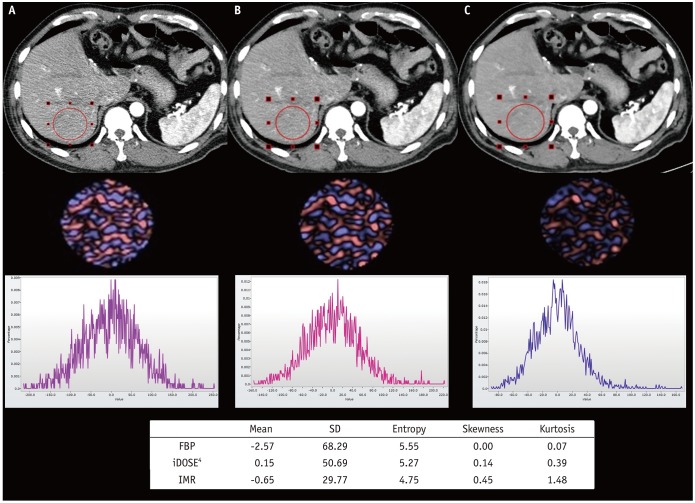
Fig. 1 Examples of filtration-histogram texture analysis process by TexRAD.First row CT images represent different reconstruction algorithms, (A) FBP, (B) iDOSE4, and (C) IMR, respectively. Second row displays processed images with fine filter value of 2. Third row displays histograms showing pixel distribution of filtered images. Table includes estimated texture feature parameters from each reconstruction algorithm. CT = computed tomography, FBP = filtered back projection, SD = standard deviation
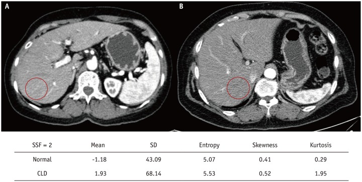
Fig. 2 Examples of normal liver group and CLD group.Arterial phase FBP image from (A) patient in normal liver group and (B) that of CLD patient. Included table shows calculated texture features from each image with filter value of 2. Among them, SD and entropy were greater in CLD patient, which were also demonstrated to be significantly different between two groups (p < 0.0001). CLD = chronic liver disease
Reference
-
1. Grootjans W, Tixier F, van der, Vriens D, Le Rest CC, Bussink J, et al. The impact of optimal respiratory gating and image noise on evaluation of intratumor heterogeneity on 18F-FDG PET imaging of lung cancer. J Nucl Med. 2016; 57:1692–1698. PMID: 27283931.
Article2. Yun BL, Cho N, Li M, Jang MH, Park SY, Kang HC, et al. Intratumoral heterogeneity of breast cancer xenograft models: texture analysis of diffusion-weighted MR imaging. Korean J Radiol. 2014; 15:591–604. PMID: 25246820.
Article3. Bashir U, Siddique MM, Mclean E, Goh V, Cook GJ. Imaging heterogeneity in lung cancer: techniques, applications, and challenges. AJR Am J Roentgenol. 2016; 207:534–543. PMID: 27305342.
Article4. Lubner MG, Smith AD, Sandrasegaran K, Sahani DV, Pickhardt PJ. CT texture analysis: definitions, applications, biologic correlates, and challenges. Radiographics. 2017; 37:1483–1503. PMID: 28898189.
Article5. Ganeshan B, Miles KA. Quantifying tumour heterogeneity with CT. Cancer Imaging. 2013; 13:140–149. PMID: 23545171.
Article6. Miles KA, Ganeshan B, Hayball MP. CT texture analysis using the filtration-histogram method: what do the measurements mean? Cancer Imaging. 2013; 13:400–406. PMID: 24061266.
Article7. Daginawala N, Li B, Buch K, Yu H, Tischler B, Qureshi MM, et al. Using texture analyses of contrast enhanced CT to assess hepatic fibrosis. Eur J Radiol. 2016; 85:511–517. PMID: 26860661.
Article8. Lubner MG, Malecki K, Kloke J, Ganeshan B, Pickhardt PJ. Texture analysis of the liver at MDCT for assessing hepatic fibrosis. Abdom Radiol (NY). 2017; 42:2069–2207. PMID: 28314916.
Article9. Ganeshan B, Abaleke S, Young RC, Chatwin CR, Miles KA. Texture analysis of non-small cell lung cancer on unenhanced computed tomography: initial evidence for a relationship with tumour glucose metabolism and stage. Cancer Imaging. 2010; 10:137–143. PMID: 20605762.
Article10. Chae HD, Park CM, Park SJ, Lee SM, Kim KG, Goo JM. Computerized texture analysis of persistent part-solid ground-glass nodules: differentiation of preinvasive lesions from invasive pulmonary adenocarcinomas. Radiology. 2014; 273:285–293. PMID: 25102296.
Article11. Miles KA, Ganeshan B, Griffiths MR, Young RC, Chatwin CR. Colorectal cancer: texture analysis of portal phase hepatic CT images as a potential marker of survival. Radiology. 2009; 250:444–452. PMID: 19164695.
Article12. Solomon J, Mileto A, Nelson RC, Roy Choudhury K, Samei E. Quantitative features of liver lesions, lung nodules, and renal stones at multi-detector row CT examinations: dependency on radiation dose and reconstruction algorithm. Radiology. 2016; 279:185–194. PMID: 26624973.
Article13. Yasaka K, Akai H, Mackin D, Court L, Moros E, Ohtomo K, et al. Precision of quantitative computed tomography texture analysis using image filtering: a phantom study for scanner variability. Medicine (Baltimore). 2017; 96:e6993. PMID: 28538408.14. Mackin D, Fave X, Zhang L, Fried D, Yang J, Taylor B, et al. Measuring computed tomography scanner variability of radiomics features. Invest Radiol. 2015; 50:757–765. PMID: 26115366.
Article15. Zhao B, Tan Y, Tsai WY, Qi J, Xie C, Lu L, et al. Reproducibility of radiomics for deciphering tumor phenotype with imaging. Sci Rep. 2016; 6:23428. PMID: 27009765.
Article16. Berrington de González A, Mahesh M, Kim KP, Bhargavan M, Lewis R, Mettler F, et al. Projected cancer risks from computed tomographic scans performed in the United States in 2007. Arch Intern Med. 2009; 169:2071–2077. PMID: 20008689.
Article17. Liu L. Model-based iterative reconstruction: a promising algorithm for today’s computed tomography imaging. J Med Imaging Radiat Sci. 2014; 45:131–136.
Article18. Yu MH, Lee JM, Yoon JH, Baek JH, Han JK, Choi BI, et al. Low tube voltage intermediate tube current liver MDCT: sinogram-affirmed iterative reconstruction algorithm for detection of hypervascular hepatocellular carcinoma. AJR Am J Roentgenol. 2013; 201:23–32. PMID: 23789655.
Article19. Yoon JH, Lee JM, Yu MH, Baek JH, Jeon JH, Hur BY, et al. Comparison of iterative model-based reconstruction versus conventional filtered back projection and hybrid iterative reconstruction techniques: lesion conspicuity and influence of body size in anthropomorphic liver phantoms. J Comput Assist Tomogr. 2014; 38:859–868. PMID: 25321625.20. Chang W, Lee JM, Lee K, Yoon JH, Yu MH, Han JK, et al. Assessment of a model-based, iterative reconstruction algorithm (MBIR) regarding image quality and dose reduction in liver computed tomography. Invest Radiol. 2013; 48:598–606. PMID: 23511193.
Article21. Kim H, Park CM, Lee M, Park SJ, Song YS, Lee JH, et al. Impact of reconstruction algorithms on CT radiomic features of pulmonary tumors: analysis of intra- and inter-reader variability and inter-reconstruction algorithm variability. PLoS One. 2016; 11:e0164924. PMID: 27741289.
Article22. Barrett HH, Myers KJ, Hoeschen C, Kupinski MA, Little MP. Task-based measures of image quality and their relation to radiation dose and patient risk. Phys Med Biol. 2015; 60:R1–R75. PMID: 25564960.
Article23. Shin JM, Kim TH, Haam S, Han K, Byun MK, Chang YS, et al. The repeatability of computed tomography lung volume measurements: comparisons in healthy subjects, patients with obstructive lung disease, and patients with restrictive lung disease. PLoS One. 2017; 12:e0182849. PMID: 28796825.
Article24. Geyer LL, Schoepf UJ, Meinel FG, Nance JW Jr, Bastarrika G, Leipsic JA, et al. State of the Art: Iterative CT Reconstruction Techniques. Radiology. 2015; 276:339–357. PMID: 26203706.
Article25. Willemink MJ, de Jong PA, Leiner T, de Heer LM, Nievelstein RA, Budde RP, et al. Iterative reconstruction techniques for computed tomography Part 1: technical principles. Eur Radiol. 2013; 23:1623–1631. PMID: 23314600.
Article26. Ng F, Ganeshan B, Kozarski R, Miles KA, Goh V. Assessment of primary colorectal cancer heterogeneity by using whole-tumor texture analysis: contrast-enhanced CT texture as a biomarker of 5-year survival. Radiology. 2013; 266:177–184. PMID: 23151829.
Article27. Elpek GÖ. Angiogenesis and liver fibrosis. World J Hepatol. 2015; 7:377–391. PMID: 25848465.
Article28. Lautt WW. Mechanism and role of intrinsic regulation of hepatic arterial blood flow: hepatic arterial buffer response. Am J Physiol. 1985; 249(5 Pt 1):G549–G556. PMID: 3904482.
Article29. Yoon JH, Lee JM, Klotz E, Jeon JH, Lee KB, Han JK, et al. Estimation of hepatic extracellular volume fraction using multiphasic liver computed tomography for hepatic fibrosis grading. Invest Radiol. 2015; 50:290–296. PMID: 25493416.
Article30. Park SB, Kim YS, Lee JB, Park HJ. Knowledge-based iterative model reconstruction (IMR) algorithm in ultralow-dose CT for evaluation of urolithiasis: evaluation of radiation dose reduction, image quality, and diagnostic performance. Abdom Imaging. 2015; 40:3137–3146. PMID: 26197735.
Article
- Full Text Links
-
- Actions
-
Cited
- CITED
-
- Close
- Share
-
- Similar articles
-
- Quantitative Image Quality and Histogram-Based Evaluations of an Iterative Reconstruction Algorithm at Low-to-Ultralow Radiation Dose Levels: A Phantom Study in Chest CT
- Quantitative Analysis of the Effect of Iterative Reconstruction Using a Phantom: Determining the Appropriate Blending Percentage
- Impact of Model-Based Iterative Reconstruction on the Correlation between Computed Tomography Quantification of a Low Lung Attenuation Area and Airway Measurements and Pulmonary Function Test Results in Normal Subjects
- Statistical Methods for Tomographic Image Reconstruction in Nuclear Medicine
- Hepatocellular Carcinoma: Texture Analysis of Preoperative Computed Tomography Images Can Provide Markers of Tumor Grade and Disease-Free Survival
