Quantitative Image Quality and Histogram-Based Evaluations of an Iterative Reconstruction Algorithm at Low-to-Ultralow Radiation Dose Levels: A Phantom Study in Chest CT
- Affiliations
-
- 1Department of Radiology and Research Institute of Radiology, University of Ulsan College of Medicine, Asan Medical Center, Seoul 05505, Korea. hwgoo@amc.seoul.kr
- KMID: 2425117
- DOI: http://doi.org/10.3348/kjr.2018.19.1.119
Abstract
OBJECTIVE
To describe the quantitative image quality and histogram-based evaluation of an iterative reconstruction (IR) algorithm in chest computed tomography (CT) scans at low-to-ultralow CT radiation dose levels.
MATERIALS AND METHODS
In an adult anthropomorphic phantom, chest CT scans were performed with 128-section dual-source CT at 70, 80, 100, 120, and 140 kVp, and the reference (3.4 mGy in volume CT Dose Index [CTDIvol]), 30%-, 60%-, and 90%-reduced radiation dose levels (2.4, 1.4, and 0.3 mGy). The CT images were reconstructed by using filtered back projection (FBP) algorithms and IR algorithm with strengths 1, 3, and 5. Image noise, signal-to-noise ratio (SNR), and contrast-to-noise ratio (CNR) were statistically compared between different dose levels, tube voltages, and reconstruction algorithms. Moreover, histograms of subtraction images before and after standardization in x- and y-axes were visually compared.
RESULTS
Compared with FBP images, IR images with strengths 1, 3, and 5 demonstrated image noise reduction up to 49.1%, SNR increase up to 100.7%, and CNR increase up to 67.3%. Noteworthy image quality degradations on IR images including a 184.9% increase in image noise, 63.0% decrease in SNR, and 51.3% decrease in CNR, and were shown between 60% and 90% reduced levels of radiation dose (p < 0.0001). Subtraction histograms between FBP and IR images showed progressively increased dispersion with increased IR strength and increased dose reduction. After standardization, the histograms appeared deviated and ragged between FBP images and IR images with strength 3 or 5, but almost normally-distributed between FBP images and IR images with strength 1.
CONCLUSION
The IR algorithm may be used to save radiation doses without substantial image quality degradation in chest CT scanning of the adult anthropomorphic phantom, down to approximately 1.4 mGy in CTDIvol (60% reduced dose).
Keyword
MeSH Terms
Figure
-

Fig. 1 Axial CT images of chest phantom obtained at reference dose level and 70 kVp.A. Axial CT image reconstructed with FBP (B30f) demonstrates locations of three regions of interest (N, lung nodule; B1, air outside anterior chest wall; B2, right posterior lung). B. Axial CT image reconstructed with sinogram-affirmed IR with strength of 5 (I30f_5) shows decrease in image noise and increase in image blurring, compared with corresponding FBP image (A). In contrast, beam-hardening artifacts caused by simulated coronary arteries in phantom remain largely unchanged between two (A, B). C. Subtraction image between two CT images (B30f-I30f_5) clearly reveals subtle differences caused by application of IR algorithm that can be difficult to recognize by visual comparison. In addition to noise pattern, distinct outlines of chest phantom are seen on subtraction image, which can explain image blurring caused by IR technique. FBP = filtered back projection, IR = iterative reconstruction
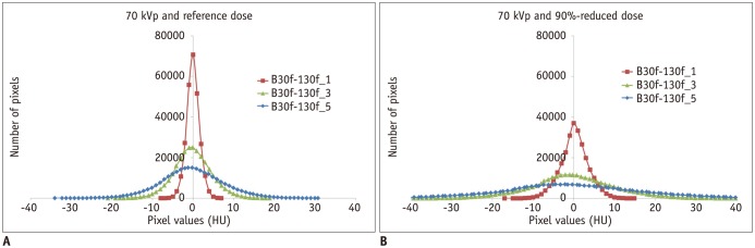
Fig. 2 Histograms of subtraction images between FBP and sinogram-affirmed IR images.Histograms of three subtraction images (B30f-I30f_1, B30f-I30f_3, and B30f-I30f_5) acquired at 70 kVp and reference radiation dose (A) and acquired at 70 kVp and 90%-reduced radiation dose (B) show gradually increased horizontal stretching with increased strength of IR algorithm. Of note, degree of their horizontal stretching is more pronounced at 90%-reduced radiation dose than at reference radiation dose for corresponding subtraction pairs. HU = Hounsfield units
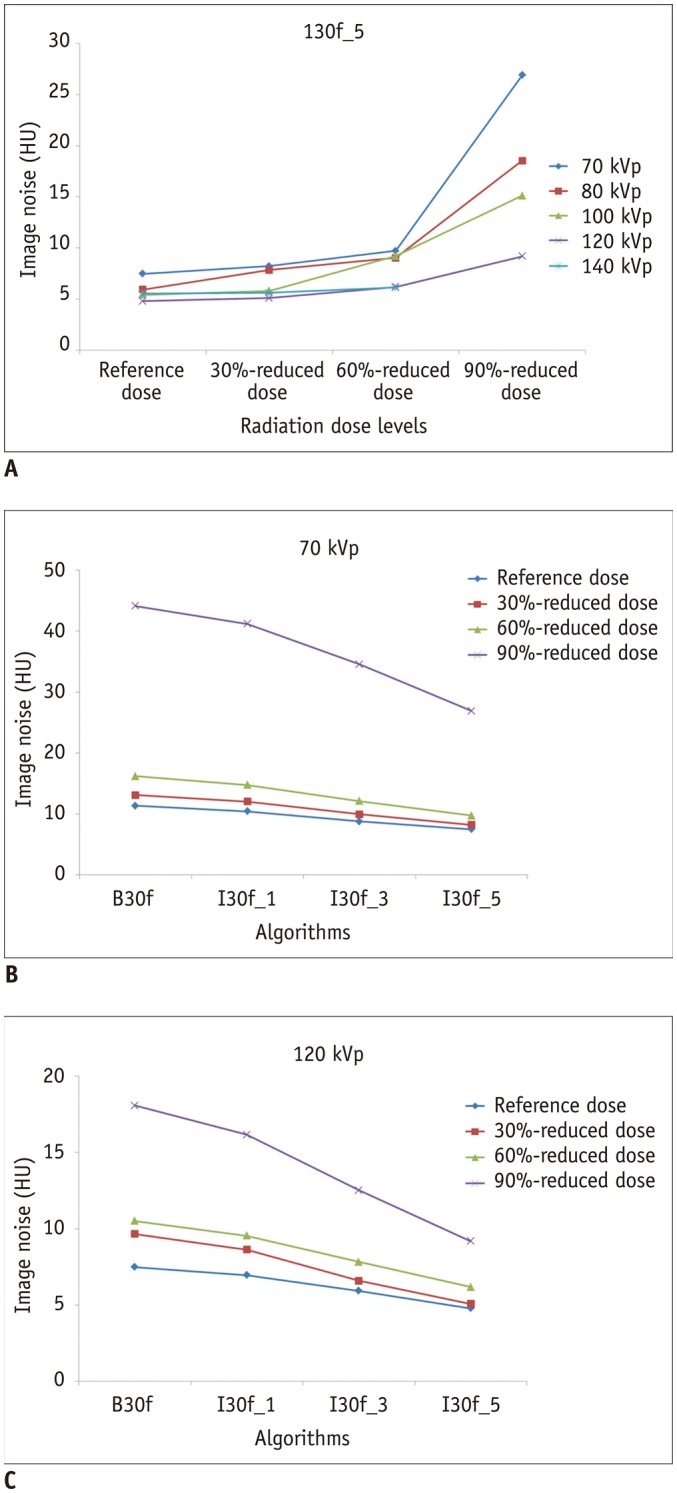
Fig. 3 Graphs demonstrating effects of radiation dose, tube voltage, and image reconstruction algorithm on image noise.A. Graph shows markedly increased image noise of CT image reconstructed with sinogram-affirmed IR with strength of 5 (I30f_5) between 60%- and 90%-reduced radiation dose levels. B, C. Graphs demonstrate greater image noise reduction with higher strength of IR algorithm at 70 kVp (B) and 120 kVp (C). Greatest image noise change is also noted between 60%- and 90%-reduced radiation dose levels at both 70 kVp (B) and 120 kVp (C).
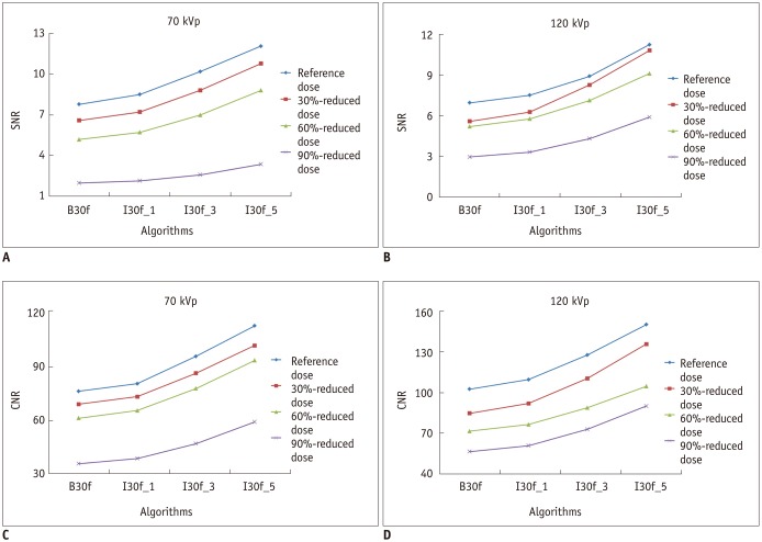
Fig. 4 Graphs demonstrating effects of radiation dose, tube voltage, and image reconstruction algorithm on SNR and CNR.A, B. Graphs show greater SNR increase with higher strength of IR algorithm at 70 kVp (A) and 120 kVp (B). Greatest SNR change is noted between 60%- and 90%-reduced radiation dose levels at both 70 kVp (A) and 120 kVp (B). C, D. Graphs show greater CNR increase with higher strength of IR algorithm at 70 kVp (C) and 120 kVp (D). In contrast to SNR, greatest SNR change is noted between 60%- and 90%-reduced radiation dose levels at 70 kVp (C) but not at 120 kVp (D). CNR = contrast-to-noise ratio, SNR = signal-to-noise ratio
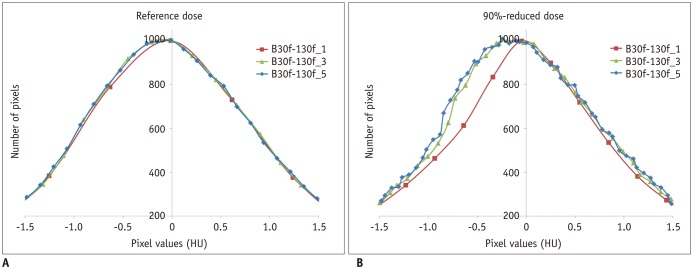
Fig. 5 Magnified standardized histograms of subtraction images between FBP and IR algorithm at 70 kVp.A. At reference radiation dose, all three histograms are almost normally distributed and slightly skewed to right. B. At 90%-reduced radiation dose, histogram shows almost normal distribution for strength 1, but histograms show deviated and ragged appearance for strengths 3 and 5.
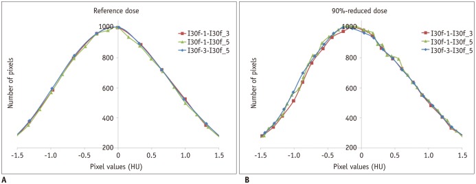
Fig. 6 Magnified standardized histograms of subtraction images between IR algorithms at 70 kVp.A. At reference radiation dose, all three histograms are almost normally distributed. B. At 90%-reduced radiation dose, all three histograms appear minimally deviated and ragged as well as slightly skewed to right for all strengths.
Cited by 3 articles
-
Semiautomatic Three-Dimensional Threshold-Based Cardiac Computed Tomography Ventricular Volumetry in Repaired Tetralogy of Fallot: Comparison with Cardiac Magnetic Resonance Imaging
Hyun Woo Goo
Korean J Radiol. 2019;20(1):102-113. doi: 10.3348/kjr.2018.0237.Application of Vendor-Neutral Iterative Reconstruction Technique to Pediatric Abdominal Computed Tomography
Woo Hyeon Lim, Young Hun Choi, Ji Eun Park, Yeon Jin Cho, Seunghyun Lee, Jung-Eun Cheon, Woo Sun Kim, In-One Kim, Jong Hyo Kim
Korean J Radiol. 2019;20(9):1358-1367. doi: 10.3348/kjr.2018.0715.User-Friendly Vendor-Specific Guideline for Pediatric Cardiothoracic Computed Tomography Provided by the Asian Society of Cardiovascular Imaging Congenital Heart Disease Study Group: Part 1. Imaging Techniques
Sun Hwa Hong, Hyun Woo Goo, Eriko Maeda, Ki Seok Choo, I-Chen Tsai,
Korean J Radiol. 2019;20(2):190-204. doi: 10.3348/kjr.2018.0571.
Reference
-
1. Brenner DJ, Hall EJ. Computed tomography--an increasing source of radiation exposure. N Engl J Med. 2007; 357:2277–2284. PMID: 18046031.2. Kalra MK, Maher MM, Toth TL, Hamberg LM, Blake MA, Shepard JA, et al. Strategies for CT radiation dose optimization. Radiology. 2004; 230:619–628. PMID: 14739312.
Article3. McCollough CH, Bruesewitz MR, Kofler JM Jr. CT dose reduction and dose management tools: overview of available options. Radiographics. 2006; 26:503–512. PMID: 16549613.
Article4. Goo HW. CT radiation dose optimization and estimation: an update for radiologists. Korean J Radiol. 2012; 13:1–11. PMID: 22247630.
Article5. Gunn ML, Kohr JR. State of the art: technologies for computed tomography dose reduction. Emerg Radiol. 2010; 17:209–218. PMID: 19936808.
Article6. Beister M, Kolditz D, Kalender WA. Iterative reconstruction methods in X-ray CT. Phys Med. 2012; 28:94–108. PMID: 22316498.
Article7. Fleischmann D, Boas FE. Computed tomography--old ideas and new technology. Eur Radiol. 2011; 21:510–517. PMID: 21249371.
Article8. Leipsic J, Heilbron BG, Hague C. Iterative reconstruction for coronary CT angiography: finding its way. Int J Cardiovasc Imaging. 2012; 28:613–620. PMID: 21359835.
Article9. Noél PB, Fingerle AA, Renger B, Münzel D, Rummeny EJ, Dobritz M. Initial performance characterization of a clinical noise-suppressing reconstruction algorithm for MDCT. AJR Am J Roentgenol. 2011; 197:1404–1409. PMID: 22109296.10. Winklehner A, Karlo C, Puippe G, Schmidt B, Flohr T, Goetti R, et al. Raw data-based iterative reconstruction in body CTA: evaluation of radiation dose saving potential. Eur Radiol. 2011; 21:2521–2526. PMID: 21822785.
Article11. Moscariello A, Takx RA, Schoepf UJ, Renker M, Zwerner PL, O'Brien TX, et al. Coronary CT angiography: image quality, diagnostic accuracy, and potential for radiation dose reduction using a novel iterative image reconstruction technique-comparison with traditional filtered back projection. Eur Radiol. 2011; 21:2130–2138. PMID: 21611758.
Article12. Kalra MK, Woisetschläger M, Dahlström N, Singh S, Lindblom M, Choy G, et al. Radiation dose reduction with sinogram affirmed iterative reconstruction technique for abdominal computed tomography. J Comput Assist Tomogr. 2012; 36:339–346. PMID: 22592621.
Article13. Tricarico F, Hlavacek AM, Schoepf UJ, Ebersberger U, Nance JW Jr, Vliegenthart R, et al. Cardiovascular CT angiography in neonates and children: image quality and potential for radiation dose reduction with iterative image reconstruction techniques. Eur Radiol. 2013; 23:1306–1315. PMID: 23207869.
Article14. Goo HW, Yang DH, Hong SJ, Yu J, Kim BJ, Seo JB, et al. Xenon ventilation CT using dual-source and dual-energy technique in children with bronchiolitis obliterans: correlation of xenon and CT density values with pulmonary function test results. Pediatr Radiol. 2010; 40:1490–1497. PMID: 20411254.
Article15. Whyne C, Hardisty M, Wu F, Skrinskas T, Clemons M, Gordon L, et al. Quantitative characterization of metastatic disease in the spine. Part II. Histogram-based analyses. Med Phys. 2007; 34:3279–3285. PMID: 17879791.
Article16. Chen X, Schott D, Song Y, Li D, Hall W, Erickson B, et al. SUF-R-50: Radiation-induced changes in CT number histogram during chemoradiation therapy for pancreatic cancer. Med phys. 2016; 43:3384.
Article17. Goo HW. Individualized volume CT dose index determined by cross-sectional area and mean density of the body to achieve uniform image noise of contrast-enhanced pediatric chest CT obtained at variable kV levels and with combined tube current modulation. Pediatr Radiol. 2011; 41:839–847. PMID: 21656275.
Article18. Schabel C, Fenchel M, Schmidt B, Flohr TG, Wuerslin C, Thomas C, et al. Clinical evaluation and potential radiation dose reduction of the novel sinogram-affirmed iterative reconstruction technique (SAFIRE) in abdominal computed tomography angiography. Acad Radiol. 2013; 20:165–172. PMID: 23099242.
Article19. Kim H, Park CM, Chae HD, Lee SM, Goo JM. Impact of radiation dose and iterative reconstruction on pulmonary nodule measurements at chest CT: a phantom study. Diagn Interv Radiol. 2015; 21:459–465. PMID: 26359871.
Article20. Higuchi K, Nagao M, Matsuo Y, Sunami S, Kamitani T, Jinnouchi M, et al. Detection of ground-glass opacities by use of hybrid iterative reconstruction (iDose) and low-dose 256-section computed tomography: a phantom study. Radiol Phys Technol. 2013; 6:299–304. PMID: 23400447.
Article21. Kalra MK, Woisetschläger M, Dahlström N, Singh S, Digumarthy S, Do S, et al. Sinogram-affirmed iterative reconstruction of low-dose chest CT: effect on image quality and radiation dose. AJR Am J Roentgenol. 2013; 201:W235–W244. PMID: 23883238.
Article22. Pourjabbar S, Singh S, Kulkarni N, Muse V, Digumarthy SR, Khawaja RD, et al. Dose reduction for chest CT: comparison of two iterative reconstruction techniques. Acta Radiol. 2015; 56:688–695. PMID: 24948790.
Article23. Baumueller S, Winklehner A, Karlo C, Goetti R, Flohr T, Russi EW, et al. Low-dose CT of the lung: potential value of iterative reconstructions. Eur Radiol. 2012; 22:2597–2606. PMID: 22699873.
Article24. Gay F, Pavia Y, Pierrat N, Lasalle S, Neuenschwander S, Brisse HJ. Dose reduction with adaptive statistical iterative reconstruction for paediatric CT: phantom study and clinical experience on chest and abdomen CT. Eur Radiol. 2014; 24:102–111. PMID: 23995879.
Article25. Lee SW, Kim Y, Shim SS, Lee JK, Lee SJ, Ryu YJ, et al. Image quality assessment of ultra low-dose chest CT using sinogram-affirmed iterative reconstruction. Eur Radiol. 2014; 24:817–826. PMID: 24442444.
Article26. Wang H, Tan B, Zhao B, Liang C, Xu Z. Raw-data-based iterative reconstruction versus filtered back projection: image quality of low-dose chest computed tomography examinations in 87 patients. Clin Imaging. 2013; 37:1024–1032. PMID: 23916244.
Article27. Hwang HJ, Seo JB, Lee HJ, Lee SM, Kim EY, Oh SY, et al. Low-dose chest computed tomography with sinogram-affirmed iterative reconstruction, iterative reconstruction in image space, and filtered back projection: studies on image quality. J Comput Assist Tomogr. 2013; 37:610–617. PMID: 23863540.28. Yang WJ, Yan FH, Liu B, Pang LF, Hou L, Zhang H, et al. Can sinogram-affirmed iterative (SAFIRE) reconstruction improve imaging quality on low-dose lung CT screening compared with traditional filtered back projection (FBP) reconstruction? J Comput Assist Tomogr. 2013; 37:301–305. PMID: 23493224.
Article29. Hu XH, Ding XF, Wu RZ, Zhang MM. Radiation dose of non-enhanced chest CT can be reduced 40% by using iterative reconstruction in image space. Clin Radiol. 2011; 66:1023–1029. PMID: 21861995.
Article30. Löve A, Olsson ML, Siemund R, Stålhammar F, Björkman-Burtscher IM, Söderberg M. Six iterative reconstruction algorithms in brain CT: a phantom study on image quality at different radiation dose levels. Br J Radiol. 2013; 86:20130388. PMID: 24049128.
Article31. Boedeker KL, Cooper VN, McNitt-Gray MF. Application of the noise power spectrum in modern diagnostic MDCT: part I. Measurement of noise power spectra and noise equivalent quanta. Phys Med Biol. 2007; 52:4027–4046. PMID: 17664593.
Article32. Shlomi D, Ben-Avi R, Balmor GR, Onn A, Peled N. Screening for lung cancer: time for large-scale screening by chest computed tomography. Eur Respir J. 2014; 44:217–238. PMID: 24525442.
Article33. Lee E, Goo HW, Lee JY. Age- and gender-specific estimates of cumulative CT dose over 5 years using real radiation dose tracking data in children. Pediatr Radiol. 2015; 45:1282–1292. PMID: 25801905.34. Infante JC, Liu Y, Rigsby CK. CT image quality in sinogram affirmed iterative reconstruction phantom study-is there a point of diminishing returns. Pediatr Radiol. 2017; 47:333–341. PMID: 27891546.
- Full Text Links
-
- Actions
-
Cited
- CITED
-
- Close
- Share
-
- Similar articles
-
- Dosimetric Effects of Low Dose 4D CT Using a Commercial Iterative Reconstruction on Dose Calculation in Radiation Treatment Planning: A Phantom Study
- Effects of Iterative Reconstruction Algorithm, Automatic Exposure Control on Image Quality, and Radiation Dose: Phantom Experiments with Coronary CT Angiography Protocols
- Low-Dose Abdominal CT Using a Deep Learning-Based Denoising Algorithm: A Comparison with CT Reconstructed with Filtered Back Projection or Iterative Reconstruction Algorithm
- Dose and Image Evaluations of Imaging for Radiotherapy
- Quantitative Analysis of the Effect of Iterative Reconstruction Using a Phantom: Determining the Appropriate Blending Percentage
