Quantitative Analysis of the Effect of Iterative Reconstruction Using a Phantom: Determining the Appropriate Blending Percentage
- Affiliations
-
- 1Department of Radiology, Research Institute of Radiological Science, Severance Hospital, Yonsei University College of Medicine, Seoul, Korea. yelv@yuhs.ac
- KMID: 2352814
- DOI: http://doi.org/10.3349/ymj.2015.56.1.253
Abstract
- PURPOSE
To investigate the optimal blending percentage of adaptive statistical iterative reconstruction (ASIR) in a reduced radiation dose while preserving a degree of image quality and texture that is similar to that of standard-dose computed tomography (CT).
MATERIALS AND METHODS
The CT performance phantom was scanned with standard and dose reduction protocols including reduced mAs or kVp. Image quality parameters including noise, spatial, and low-contrast resolution, as well as image texture, were quantitatively evaluated after applying various blending percentages of ASIR. The optimal blending percentage of ASIR that preserved image quality and texture compared to standard dose CT was investigated in each radiation dose reduction protocol.
RESULTS
As the percentage of ASIR increased, noise and spatial-resolution decreased, whereas low-contrast resolution increased. In the texture analysis, an increasing percentage of ASIR resulted in an increase of angular second moment, inverse difference moment, and correlation and in a decrease of contrast and entropy. The 20% and 40% dose reduction protocols with 20% and 40% ASIR blending, respectively, resulted in an optimal quality of images with preservation of the image texture.
CONCLUSION
Blending the 40% ASIR to the 40% reduced tube-current product can maximize radiation dose reduction and preserve adequate image quality and texture.
Keyword
MeSH Terms
Figure
-
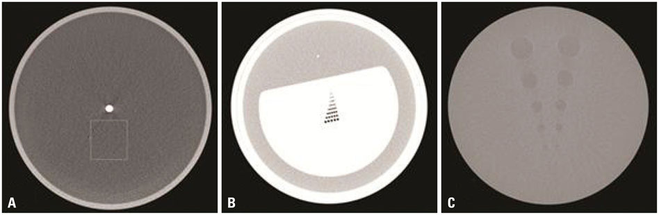
Fig. 1 American Association of Physicists in Medicine (AAPM) CT performance phantom. Selected blocks from the AAPM phantom were used to quantify noise (A), spatial resolution (B), low-contrast resolution, and texture (C).
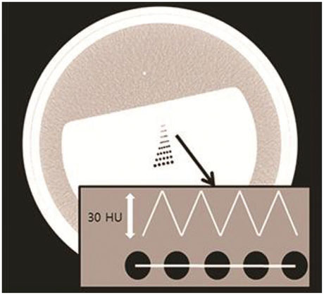
Fig. 2 Schematic graph of a HU graph drawn by the linear HU difference between five holes of the same size. HU, Hounsfield unit.

Fig. 3 Noise (standard deviation of CT numbers) of different acquisition protocols according to increasing ASIR percentage. The reference line shows the acceptable noise value of 7. If the value is <7, the quality of image is considered acceptable. ASIR, adaptive statistical iterative reconstruction.
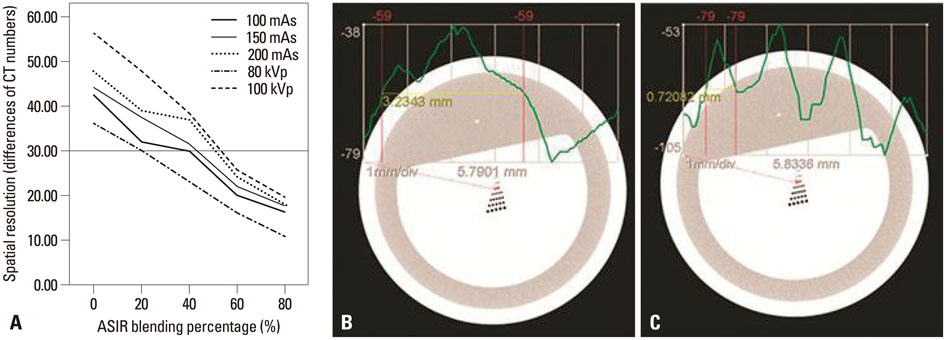
Fig. 4 Average HU differences between the peaks and valleys are shown according to increasing ASIR percentage. A difference of at least 30 HU needed to be met for optimal quality in terms of spatial resolution (A). Two graphs of the 150-mAs acquisition protocol with ASIR 100% (B) and 40% (C) are shown. HU, Hounsfield unit; ASIR, adaptive statistical iterative reconstruction.

Fig. 5 Contrast to noise ratio that was obtained to quantify low-contrast resolution according to an increase in the percentage of ASIR. The reference line indicates a value of 1.63, which was defined as the lowest acceptable limit. ASIR, adaptive statistical iterative reconstruction.
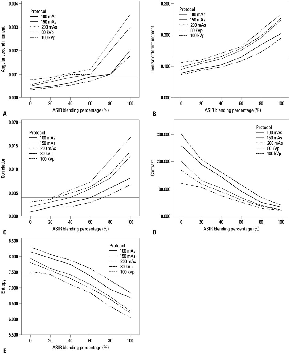
Fig. 6 Texture analysis graphs showing five different texture features according to an increase in the percentage of ASIR: angular second moment (A), inverse different moment (B), correlation (C), contrast (D), and entropy (E). The reference line of each graph shows the value of standard images.
Cited by 1 articles
-
Image Quality and Radiation Dose in CT Venography Using Model-Based Iterative Reconstruction at 80 kVp versus Adaptive Statistical Iterative Reconstruction-V at 70 kVp
Chankue Park, Ki Seok Choo, Jin Hyeok Kim, Kyung Jin Nam, Ji Won Lee, Jin You Kim
Korean J Radiol. 2019;20(7):1167-1175. doi: 10.3348/kjr.2018.0897.
Reference
-
1. Silva AC, Lawder HJ, Hara A, Kujak J, Pavlicek W. Innovations in CT dose reduction strategy: application of the adaptive statistical iterative reconstruction algorithm. AJR Am J Roentgenol. 2010; 194:191–199.
Article2. Brenner DJ, Hall EJ. Computed tomography--an increasing source of radiation exposure. N Engl J Med. 2007; 357:2277–2284.
Article3. Singh S, Kalra MK, Gilman MD, Hsieh J, Pien HH, Digumarthy SR, et al. Adaptive statistical iterative reconstruction technique for radiation dose reduction in chest CT: a pilot study. Radiology. 2011; 259:565–573.
Article4. Brady SL, Yee BS, Kaufman RA. Characterization of adaptive statistical iterative reconstruction algorithm for dose reduction in CT: A pediatric oncology perspective. Med Phys. 2012; 39:5520–5531.
Article5. García G, Maiora J, Tapia A, De Blas M. Evaluation of texture for classification of abdominal aortic aneurysm after endovascular repair. J Digit Imaging. 2012; 25:369–376.
Article6. Kato H, Kanematsu M, Zhang X, Saio M, Kondo H, Goshima S, et al. Computer-aided diagnosis of hepatic fibrosis: preliminary evaluation of MRI texture analysis using the finite difference method and an artificial neural network. AJR Am J Roentgenol. 2007; 189:117–122.
Article7. Yanagawa M, Honda O, Yoshida S, Kikuyama A, Inoue A, Sumikawa H, et al. Adaptive statistical iterative reconstruction technique for pulmonary CT: image quality of the cadaveric lung on standard- and reduced-dose CT. Acad Radiol. 2010; 17:1259–1266.
Article8. Prakash P, Kalra MK, Kambadakone AK, Pien H, Hsieh J, Blake MA, et al. Reducing abdominal CT radiation dose with adaptive statistical iterative reconstruction technique. Invest Radiol. 2010; 45:202–210.
Article9. Mendler MH, Bouillet P, Le Sidaner A, Lavoine E, Labrousse F, Sautereau D, et al. Dual-energy CT in the diagnosis and quantification of fatty liver: limited clinical value in comparison to ultrasound scan and single-energy CT, with special reference to iron overload. J Hepatol. 1998; 28:785–794.
Article10. Prakash P, Kalra MK, Digumarthy SR, Hsieh J, Pien H, Singh S, et al. Radiation dose reduction with chest computed tomography using adaptive statistical iterative reconstruction technique: initial experience. J Comput Assist Tomogr. 2010; 34:40–45.
Article11. Judy P, Balter S, Bassano D, McCullough E, Payne J, Rothenberg L. AAPM report No. 1 phantoms of performance evaluation and quality assurance of CT scanner. Chicago, IL: American Association of Physicists in Medicine;1977.12. Park HJ, Jung SE, Lee YJ, Cho WI, Do KH, Kim SH, et al. The relationship between subjective and objective parameters in CT phantom image evaluation. Korean J Radiol. 2009; 10:490–495.
Article13. Haralick RM, Shanmugam K, Dinstein IH. Textural features for image classification. IEEE Trans Syst Man Cybern B Cybern. 1973; 3:610–621.
Article14. May MS, Wüst W, Brand M, Stahl C, Allmendinger T, Schmidt B, et al. Dose reduction in abdominal computed tomography: intraindividual comparison of image quality of full-dose standard and half-dose iterative reconstructions with dual-source computed tomography. Invest Radiol. 2011; 46:465–470.
- Full Text Links
-
- Actions
-
Cited
- CITED
-
- Close
- Share
-
- Similar articles
-
- Dosimetric Effects of Low Dose 4D CT Using a Commercial Iterative Reconstruction on Dose Calculation in Radiation Treatment Planning: A Phantom Study
- Effects of Iterative Reconstruction Algorithm, Automatic Exposure Control on Image Quality, and Radiation Dose: Phantom Experiments with Coronary CT Angiography Protocols
- The Impact of Iterative Reconstruction in Low-Dose Computed Tomography on the Evaluation of Diffuse Interstitial Lung Disease
- Quantitative Image Quality and Histogram-Based Evaluations of an Iterative Reconstruction Algorithm at Low-to-Ultralow Radiation Dose Levels: A Phantom Study in Chest CT
- Adaptive Iterative Dose Reduction Algorithm in CT: Effect on Image Quality Compared with Filtered Back Projection in Body Phantoms of Different Sizes
