Stress distribution of molars restored with minimal invasive and conventional technique: a 3-D finite element analysis
- Affiliations
-
- 1Department of Pedodontics, School of Dentistry, Chonnam National University, Gwangju, Republic of Korea.
- 2Department of Bio and Brain Engineering, KAIST, Daejon, Republic of Korea.
- 3Department of Prosthodontics, School of Dentistry, Chonnam National University, Gwangju, Republic of Korea. yhsdent@jnu.ac.kr
- KMID: 2432366
- DOI: http://doi.org/10.14368/jdras.2018.34.4.297
Abstract
- PURPOSE
This study aimed to analyze stress distribution and maximum von Mises stress generated in intracoronal restorations and in tooth structures of mandibular molars with various types of cavity designs and materials.
MATERIALS AND METHODS
Threedimensional solid models of mandible molar such as O inlay cavity with composite and gold (OR-C, OG-C), MO inlay cavity with composite and gold (MR-C, MG-C), and minimal invasive cavity on occlusal and proximal surfaces (OR-M, MR-M) were designed. To simulate masticatory force, static axial load with total force of 200 N was applied on the tooth at 10 occlusal contact points. A finite element analysis was performed to predict stress distribution generated by occlusal loading.
RESULTS
Restorations with minimal cavity design generated significantly lower values of von Mises stress (OR-M model: 26.8 MPa; MR-M model: 72.7 MPa) compared to those with conventional cavity design (341.9 MPa to 397.2 MPa). In tooth structure, magnitudes of maximum von Mises stresses were similar among models with conventional design (372.8 - 412.9 MPa) and models with minimal cavity design (361.1 - 384.4 MPa).
CONCLUSION
Minimal invasive models generated smaller maximum von Mises stresses within restorations. Within the enamel, similar maximum von Mises stresses were observed for models with minimal cavity design and those with conventional design.
Keyword
Figure
-

Fig. 1 (A) Stereolithography (STL) scan image of mandibular first molar. (B) 3D CAD model of pulp, dentin, and enamel were generated and assembled.
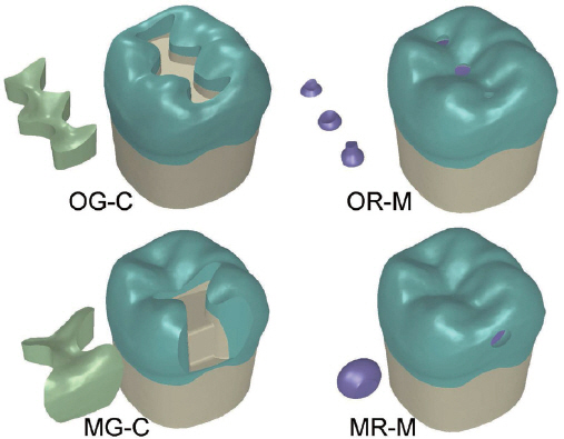
Fig. 2 Two conventional inlay models of O cavity (CGC), MO cavity (MG-C), and two minimal invasive designs for occlusal caries (OR-M) and proximal caries (MR-M) models were made.

Fig. 3 A total amount of 200 N axial load was applied at 10 occlusal points: 5 points in buccal cusp area and 5 points in marginal ridges and central fossa area (model OG-C). Solid model with restoration, enamel, dentin, and pulp chamber of the mandibular molar was meshed with tetrahedral elements. Bottom of the model was fixed in all directions as a boundary condition.
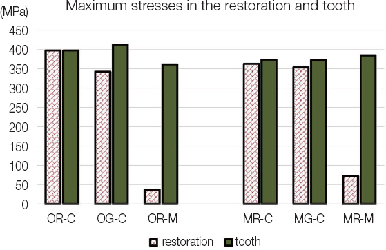
Fig. 4 Minimal invasive cavity designs (OR-M, MR-M) produced very small maximum von Mises stress magnitude compared to conventional inlay designs in the restoration. There were no significant differences in maximum stress magnitudes within the abutment tooth among models.
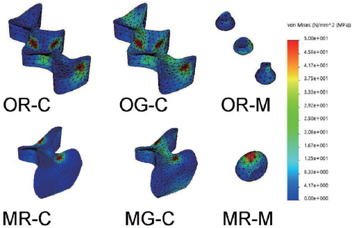
Fig. 5 Von Mises stress distribution generated by occlusal loading in the restoration of each experimental model. Models with composite (OR-C, MR-C) showed stress concentration at the loading area. Models with gold alloy (OG-C, MG-C) showed widely distributed stresses within the restorations.
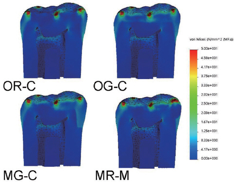
Fig. 6 Von Mises stress distribution by occlusal loading in the restoration and the abutment tooth with M-D cross-sectional view. Models of OG-C and MG-M produced well distributed stress inside the gold restoration.
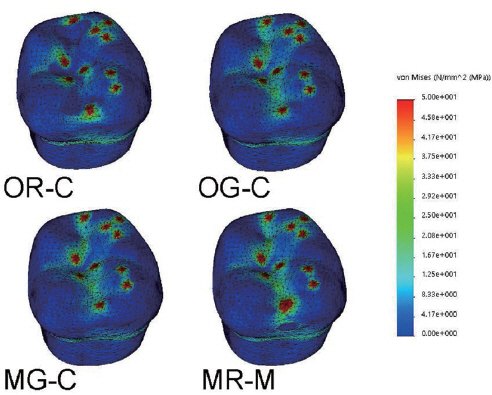
Fig. 7 Cavosurface margin between resin restorations (OR-C, MR-M) and tooth structures showed significant difference in stress gradient and distribution. Peak stress was observed at the occlusal loading area and cemento-enamel junction in all models.
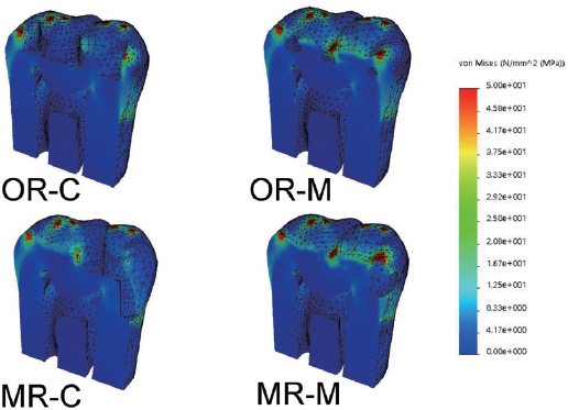
Fig. 8 Von Mises stress distribution by occlusal loading in the abutment tooth with M-D cross-sectional view. Note stress concentration was observed around the occlusal contact area, pulp horns, and cemento-enamel junction.
Reference
-
References
1. Sabbagh J, McConnell RJ, McConnell MC. Posterior composites: Update on cavities and filling techniques. J Dent. 2017; 57:86–90. DOI: 10.1016/j.jdent.2016.11.010. PMID: 27889605.2. Tyas MJ, Anusavice KJ, Frencken JE, Mount GJ. Minimal intervention dentistry - a review. FDI Commission Project 1-97. Int Dent J. 2000; 50:1–12. DOI: 10.1111/j.1875-595X.2000.tb00540.x. PMID: 10945174.3. Lynch CD, Opdam NJ, Hickel R, Brunton PA, Gurgan S, Kakaboura A, Shearer AC, Vanherle G, Wilson NH. Guidance on posterior resin composites: Academy of Operative Dentistry - European Section. J Dent. 2014; 42:377–83. DOI: 10.1016/j.jdent.2014.01.009. PMID: 24462699.4. Banerjee A. Minimal intervention dentistry: part 7. Minimally invasive operative caries management: rationale and techniques. Br Dent J. 2013; 214:107–11. DOI: 10.1038/sj.bdj.2013.106. PMID: 23392023.5. Ericson D, Kidd E, McComb D, Mjör I, Noack MJ. Minimally Invasive Dentistry - concepts and techniques in cariology. Oral Health Prev Dent. 2003; 1:59–72. PMID: 15643750.6. Tassery H, Levallois B, Terrer E, Manton DJ, Otsuki M, Koubi S, Gugnani N, Panayotov I, Jacquot B, Cuisinier F, Rechmann P. Use of new minimum intervention dentistry technologies in caries management. Aust Dent J. 2013; 58(Suppl 1):40–59. DOI: 10.1111/adj.12049. PMID: 23721337.7. Casagrande L, Laske M, Bronkhorst EM, Huysmans MCDNJM, Opdam NJM. Repair may increase survival of direct posterior restorations - A practice based study. J Dent. 2017; 64:30–6. DOI: 10.1016/j.jdent.2017.06.002. PMID: 28602850.8. Staehle HJ, Wohlrab T, Saure D, Wolff D, Frese C. A 6.5-year clinical follow-up of direct resin composite buildups in the posterior dentition: Introduction of a new minimally invasive restorative method. J Dent. 2015; 43:1211–7. DOI: 10.1016/j.jdent.2015.07.007. PMID: 26165864.9. White JM, Eakle WS. Rationale and treatment approach in minimally invasive dentistry. J Am Dent Assoc. 2000; 131:13S–9S. DOI: 10.14219/jada.archive.2000.0394. PMID: 10860340.10. Abu-Hanna AA, Mjör IA. Resin composite reinforcement of undermined enamel. Oper Dent. 2004; 29:234–7. PMID: 15088737.11. Eidelman E. Composite resin support of undermined enamel in amalgam restorations. Pediatr Dent. 1999; 21:118–20. PMID: 10197337.12. Fonseca RB, Fernandes-Neto AJ, Correr-Sobrinho L, Soares CJ. The influence of cavity preparation design on fracture strength and mode of fracture of laboratory processed composite resin restorations. J Prosthet Dent. 2007; 98:277–84. DOI: 10.1016/S0022-3913(07)60101-2. PMID: 17936127.13. Yang H, Park C, Shin JH, Yun KD, Lim HP, Park SW, Chung H. Stress distribution in premolars restored with inlays or onlays: 3D finite element analysis. J Adv Prosthodont. 2018; 10:184–90. DOI: 10.4047/jap.2018.10.3.184. PMID: 29930787. PMCID: PMC6004358.14. St-Georges AJ, Sturdevant JR, Swift EJ Jr, Thompson JY. Fracture resistance of prepared teeth restored with bonded inlay restorations. J Prosthet Dent. 2003; 89:551–7. DOI: 10.1016/S0022-3913(03)00173-2. PMID: 12815348.15. Costa A, Xavier T, Noritomi P, Saavedra G, Borges A. The influence of elastic modulus of inlay materials on stress distribution and fracture of premolars. Oper Dent. 2014; 39:E160–70. DOI: 10.2341/13-092-L. PMID: 24967990.16. Yamanel K, Caglar A, Gülsahi K, Ozden UA. Effects of different ceramic and composite materials on stress distribution in inlay and onlay cavities: 3-D finite element analysis. Dent Mater J. 2009; 28:661–70. DOI: 10.4012/dmj.28.661. PMID: 20019416.17. Latino C, Troendle K, Summitt JB. Support of undermined occlusal enamel provided by restorative materials. Quintessence Int. 2001; 32:287–91. PMID: 12066648.18. Soares CJ, Martins LR, Pfeifer JM, Giannini M. Fracture resistance of teeth restored with indirectcomposite and ceramic inlay systems. Quintessence Int. 2004; 35:281–6. PMID: 15119713.19. Dejak B, Mlotkowski A. Three-dimensional finite element analysis of strength and adhesion of composite resin versus ceramic inlays in molars. J Prosthet Dent. 2008; 99:131–40. DOI: 10.1016/S0022-3913(08)60029-3. PMID: 18262014.20. Jiang W, Bo H, Yongchun G, LongXing N. Stress distribution in molars restored with inlays or onlays with or without endodontic treatment: a threedimensional finite element analysis. J Prosthet Dent. 2010; 103:6–12. DOI: 10.1016/S0022-3913(09)60206-7. PMID: 20105674.21. Zarone F, Sorrentino R, Apicella D, Valentino B, Ferrari M, Aversa R, Apicella A. Evaluation of the biomechanical behavior of maxillary central incisors restored by means of endocrowns compared to a natural tooth: a 3D static linear finite elements analysis. Dent Mater. 2006; 22:1035–44. DOI: 10.1016/j.dental.2005.11.034. PMID: 16406084.22. Guven S, Akdogan M, Oz C, Dogan MS, Unal M, Unal S, Sahbaz C. Three-dimensional finite-element analysis of two ceramic inlay restorations with different cavity designs. Biotechnol Biotechnol Equip. 2015; 29:579–85. DOI: 10.1080/13102818.2015.1015445.23. Magne P. Efficient 3D finite element analysis of dental restorative procedures using micro-CT data. Dent Mater. 2007; 23:539–48. DOI: 10.1016/j.dental.2006.03.013. PMID: 16730058.24. Heo KH, Lim YJ, Kim MJ, Kwon HB. Three-dimensional finite element analysis of the splinted implant prosthesis in a reconstructed mandible. J Adv Prosthodont. 2018; 10:138–46. DOI: 10.4047/jap.2018.10.2.138. PMID: 29713435. PMCID: PMC5917106.25. Zelic K, Vukicevic A, Jovicic G, Aleksandrovic S, Filipovic N, Djuric M. Mechanical weakening of devitalized teeth: three-dimensional Finite Element Analysis and prediction of tooth fracture. Int Endod J. 2015; 48:850–63. DOI: 10.1111/iej.12381. PMID: 25243348.26. Wayne JS, Chande R, Porter HC, Janus C. Effect of restoration volume on stresses in a mandibular molar: a finite element study. J Prosthet Dent. 2014; 112:925–31. DOI: 10.1016/j.prosdent.2014.01.006. PMID: 24726589.27. Ausiello P, Franciosa P, Martorelli M, Watts DC. Numerical fatigue 3D-FE modeling of indirect composite-restored posterior teeth. Dent Mater. 2011; 27:423–30. DOI: 10.1016/j.dental.2010.12.001. PMID: 21227484.28. Thompson MC, Thompson KM, Swain M. The allceramic, inlay supported fixed partial denture. Part 1. Ceramic inlay preparation design: a literature review. Aust Dent J. 2010; 55:120–7. DOI: 10.1111/j.1834-7819.2010.01214.x. PMID: 20604751.
- Full Text Links
-
- Actions
-
Cited
- CITED
-
- Close
- Share
-
- Similar articles
-
- The effect of restorative materials on the stress distribution of class V composite resin restorations: a 3D finite element investigation
- Effect of the marginal position of prosthesis on stress distribution of teeth with abfraction lesion using finite element analysis
- A three-dimensional Finite element analysis for initial stress of maxillary incisors during activation of upper utility arch wire
- Stress distribution of endodontically treated maxillary second premolars restored with different methods: Three-dimensional finite element analysis
- The influence of combining composite resins with different elastic modulus on the stress distribution of Class V restoration: a three-dimensional finite element study
