Retropharyngeal Tenosynovial Giant Cell Tumor Misdiagnosed as Oropharyngeal Cancer: a Case Report
- Affiliations
-
- 1Department of Radiology, Dankook University Hospital, Cheonan, Korea. minipacs@dkuh.co.kr
- KMID: 2431114
- DOI: http://doi.org/10.13104/imri.2018.22.4.272
Abstract
- Extra-articular tenosynovial giant cell tumor (TS-GCT) in retropharyngeal space is a rare case. We found only two case reports in the literature, in which one was located in retropharynx or prevertebral space of the cervical spine. We describe a rare case of TS-GCT in the retropharynx, which was initially misdiagnosed as oropharyngeal cancer. Furthermore, we want to assure that extraarticular diffuse type TS-GCT should be considered in the differential diagnosis of lesions showing low signal intensity in MRI scan.
Keyword
MeSH Terms
Figure
-
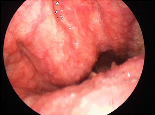
Fig. 1 Laryngoscopy reveals a smooth margined bulging contour mass lesion in the posterior oropharynx.

Fig. 2 Cervical spine CT scan bone setting axial (a), sagittal (b) and soft tissue setting with enhancement (c). (a, b) The axial/reformatted sagittal bone algorithm shows bony lytic lesion with partially very thin sclerotic margin involving C2 odontoid process and vertebral body, abutting with the inferior margin of the anterior atlantoaxial (AA) joint. (c) Sagittal contrast-enhanced CT scan shows a lytic, heterogeneously enhancing soft-tissue density mass involving the C2 extending to prevertebral space, as expected from a mass originating in the prevertebral space.
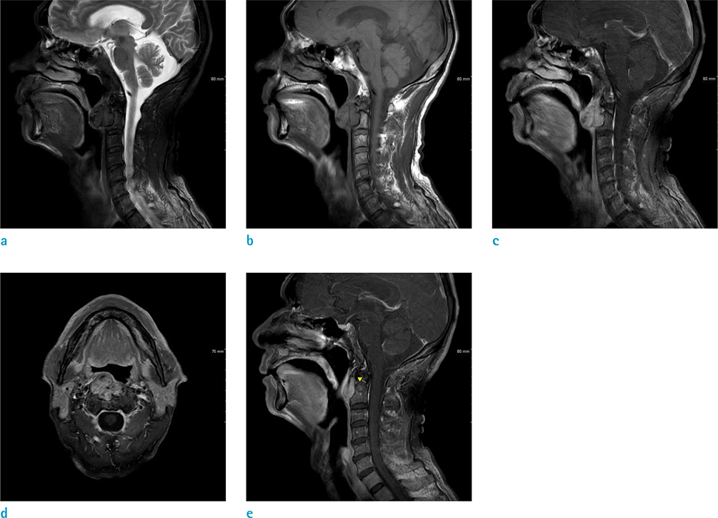
Fig. 3 T1 sagittal (a), T2 fat suppression sagittal (b), T1 enhancement sagittal (c) (e), axial (d). (a) Sagittal T1 weighted image shows heterogeneously intermediate signal intensity mass involving the odontoid process and vertebral body of C2 and abutting with the inferior margin of the anterior atlantoaxial (AA) joint. (b) Sagittal T2 weighted image with fat-suppression shows markedly hypointense mass. (c) T1 weighted image with contrast enhancement sagittal view, (d) axial view shows heterogeneous enhancement and no evidence of perilesional extension. (e) Enhanced T1 weighted image sagittal view shows the mass abutting with the inferior border of the atlantoaxial joint but nearly not involving the joint.
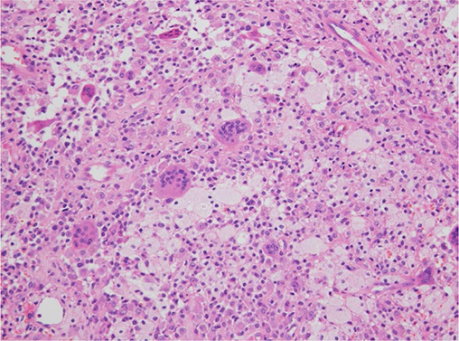
Fig. 4 The pathologic finding shows numerous foamy histiocytes, scattered multinucleated giant cells, consistent with diffuse type tenosynovial giant cell tumor (Hematoxylin & Eosin staining, × 200).
Reference
-
1. Koontz NA, Quigley EP, Witt BL, Sanders RK, Shah LM. Pigmented villonodular synovitis of the cervical spine: case report and review of the literature. BJR Case Rep. 2016; 2:20150264.
Article2. Rateb K, Hassen BG, Leila A, Faten F, Med Samir D. Giant cell tumor of soft tissues: a case report of extra-articular diffuse-type giant cell tumor of the quadriceps. Int J Surg Case Rep. 2017; 31:245–249.
Article3. Paulino AF, Spiro RH, O'Malley B, Huvos AG. Giant cell tumour of the retropharynx. Histopathology. 1998; 33:344–348.
Article4. Jaffe HL, Lichtenstein L, Sutro CJ. Pigmented villonodular synovitis, bursitis and tenosynovitis. A discussion of synovial and bursal equivalents of the tenosynovial lesion commonly denoted as xanthorna, xanthogranuloma, giant cell tumor or myeloplaxoma of the tendon sheath, with some consideration of this tendon sheath lesion itself. Arch Pathol. 1941; 31:731–765.5. Savvidou OD, Mavrogenis AF, Sakellariou VI, Chloros GD, Sarlikiotis T, Papagelopoulos PJ. Extra-articular diffuse giant cell tumor of the tendon sheath: a report of 2 cases. Arch Bone Jt Surg. 2016; 4:273–276.6. Lucas DR. Tenosynovial giant cell tumor: case report and review. Arch Pathol Lab Med. 2012; 136:901–906.
Article7. Ravi V, Wang W, Araujo DM, et al. Imatinib in the treatment of tenosynovial giant-cell tumor and pigmented villonodular synovitis. J Clin Oncol. 2010; 28(15s):10011.
Article8. Kuhnen C, Muller KM, Rabstein S, Kasprzynski A, Herter P. Tenosynovial giant cell tumor. Pathologe. 2005; 26:96–110.9. Dingle SR, Flynn JC, Flynn JC Jr, Stewart G. Giant-cell tumor of the tendon sheath involving the cervical spine. A case report. J Bone Joint Surg Am. 2002; 84-A:1664–1667.10. Blay JY, El Sayadi H, Thiesse P, Garret J, Ray-Coquard I. Complete response to imatinib in relapsing pigmented villonodular synovitis/tenosynovial giant cell tumor (PVNS/TGCT). Ann Oncol. 2008; 19:821–822.
Article
- Full Text Links
-
- Actions
-
Cited
- CITED
-
- Close
- Share
-
- Similar articles
-
- Tenosynovial Giant-cell Tumor of the Lumbar Spine
- Tenosynovial Giant Cell Tumor Showing Severe Bone Erosion in the Finger: Case Report and Review of the Imaging Findings and Their Significance
- A Tenosynovial Giant Cell Tumor Arising from Femoral Attachment of the Anterior Cruciate Ligament
- Tenosynovial Giant Cell Tumor of the Temporomandibular Joint
- Tenosynovial giant cell tumor of finger, localized type: a case report
