A Tenosynovial Giant Cell Tumor Arising from Femoral Attachment of the Anterior Cruciate Ligament
- Affiliations
-
- 1Department of Orthopedic Surgery, Chonbuk National University Hospital, Jeonju, Korea. wsi1205@naver.com
- KMID: 1784674
- DOI: http://doi.org/10.4055/cios.2014.6.2.242
Abstract
- The localized type of tenosynovial giant cell tumor usually occurs on the palmar side of fingers and toes. Tenosynovial giant cell tumors of the tendon sheath are rarely intra-articular. We report a giant cell tumor of the tendon sheath arising from femoral attachment of the anterior cruciate ligament and its treatment with arthroscopy in a 28-year-old man.
MeSH Terms
Figure
-
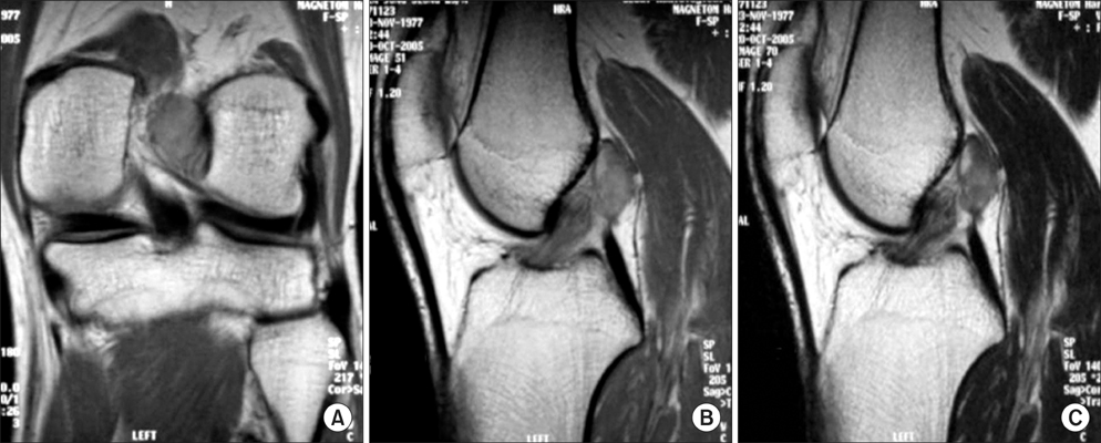
Fig. 1 (A, B) Coronal and sagittal T1-weighted images demonstrated that the soft tissue mass was isointense to muscle and obscured the anterior cruciate ligament. (C) T2-weighted sagittal magnetic resonance image showed heterogenous, intermediate to low signal intensity, which was slightly higher than that of skeletal muscle.
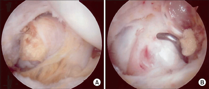
Fig. 2 (A) The mass was located behind the anterior cruciate ligament, close to the femoral attachment site. It was round in shape, measuring about 20 mm × 11 mm in diameter with a reddish-brown color. (B) It was excised with the use of a motorized instrument and basket forceps by piecemeal.

Fig. 3 Microscopic appearance of the tumor. (A) The basic cellular composition of the tumor was well-defined polygonal mononuclear cells with a scanty, faintly eosinophilic cytoplasm. In some areas, there were bands or sheets of amorphous collagen (H&E, ×100). (B) Foci of xanthoma cells with foamy cytoplasm and vacuoles were present, accompanied by branching capillaries. Multinucleated giant cells had abundant eosinophilic cytoplasm and contained eight or more nuclei (H&E, ×400).
Reference
-
1. Weiss SW, Goldblum JR. Enzinger and Weiss's soft tissue tumors. 4th ed. Philadelphia, PA: Mosby;2001. p. 1037–1062.2. Kuhnen C, Muller KM, Rabstein S, Kasprzynski A, Herter P. Tenosynovial giant cell tumor. Pathologe. 2005; 26(2):96–110.3. Otsuka Y, Mizuta H, Nakamura E, Kudo S, Inoue S, Takagi K. Tenosynovial giant-cell tumor arising from the anterior cruciate ligament of the knee. Arthroscopy. 1996; 12(4):496–499.4. Jones FE, Soule EH, Coventry MB. Fibrous xanthoma of synovium (giant-cell tumor of tendon sheath, pigmented nodular synovitis): a study of one hundred and eighteen cases. J Bone Joint Surg Am. 1969; 51(1):76–86.5. Miettinen MM. Diagnostic soft tissue pathology. Philadelphia, PA: Churchill Livingstone;2003. p. 489–502.6. Wood GS, Beckstead JH, Medeiros LJ, Kempson RL, Warnke RA. The cells of giant cell tumor of tendon sheath resemble osteoclasts. Am J Surg Pathol. 1988; 12(6):444–452.
- Full Text Links
-
- Actions
-
Cited
- CITED
-
- Close
- Share
-
- Similar articles
-
- A Tenosynovial Giant Cell Tumor Arising from Posterior Cruciate Ligament of Knee Joint: A Case Report
- Avulsion of the Femoral Attachment of Anterior Cruciate Ligament Associated with Ipsilateral Femoral Shaft Fracture in Skeletally Mature Patient: A Case Report
- Tenosynovial Giant-cell Tumor of the Lumbar Spine
- A Clinical Study on Ligamentous Injuries of the Knee
- Open anterior cruciate ligament reconstruction using inside-out femoral drilling
