Tenosynovial Giant Cell Tumor Showing Severe Bone Erosion in the Finger: Case Report and Review of the Imaging Findings and Their Significance
- Affiliations
-
- 1Department of Radiology, Jeju National University Hospital, Jeju-si, Korea. we1977@naver.com
- 2Department of Pathology, Jeju National University Hospital, Jeju-si, Korea.
- KMID: 2327426
- DOI: http://doi.org/10.13104/imri.2016.20.2.127
Abstract
- We report a case of tenosynovial giant cell tumor with severe bone erosion in the right fifth finger of a 46-year-old man. Throughout this case review, we describe the imaging findings of tenosynovial giant cell tumor with severe bone erosion and review the literatures regarding osseous lesions caused by tenosynovial giant cell tumor and their significance related to the differential diagnosis and patient treatment.
Keyword
MeSH Terms
Figure
-
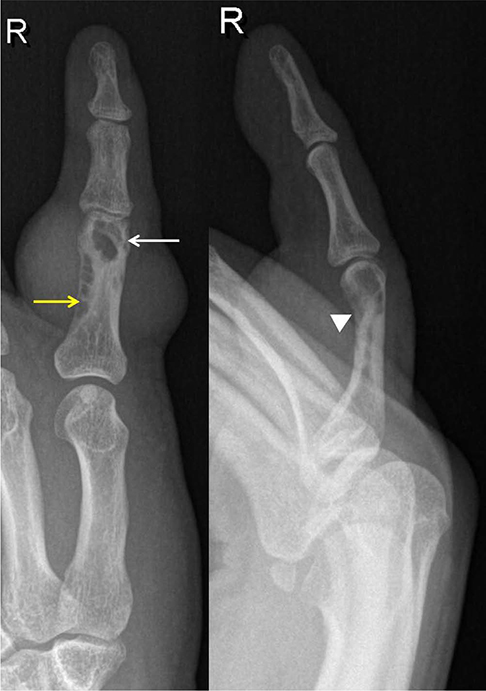
Fig. 1 Plain radiographs show a soft-tissue density showing bulging contour in the proximal phalanx level of the right fifth finger as well as intramedullary and cortical cystic lesions (white arrow) with trabeculation (yellow arrow) and cortical erosion (arrowhead) in the proximal phalanx.
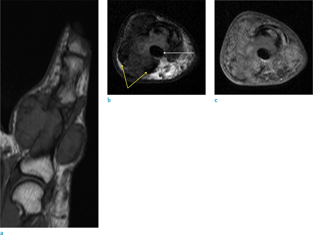
Fig. 2 Coronal T1-weighted image (a) reveals a soft-tissue tumor associated with an intraosseous lesion. On the axial T2-weighted image (b), the tumor is seen to grow surrounding the flexor digitorum tendon (white arrow) and show severe bone erosion. There is dark signal intensity (yellow arrows) in the periphery of the mass. On axial gadolinium-enhanced T1-weighted image with fat saturation (c), enhancement of the tumor is seen.

Fig. 3 Bone scan shows mild uptake (arrow) in the proximal phalanx of the right fifth finger.
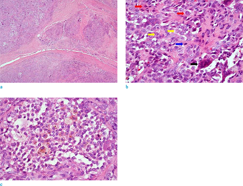
Fig. 4 Photomicrograph shows the multinodular growth pattern of the tumor with fibrous septa and stromal fibrosis (a, Hematoxylin & Eosin, × 40). The tumor is composed of a polymorphous population of mononuclear stromal cells (yellow arrows) with small round, spindle, rentiform nuclei, epithelioid macrophages (red arrows) with abundant eosinophilic cytoplasm, vesicular nuclei and osteoclast-like giant cells (black arrow). Mitotic figure is also noted (blue arrow) (b, Hematoxylin & Eosin, × 400). Sheets and clusters of xanthoma cells are frequently observed. Xanthoma cells (foamy macrophages) have copious, vacuolated cytoplasm and central nuclei. Siderophages (hemosiderin-laden macrophages) are also noted. (c, Hematoxylin & Eosin, × 400).
Reference
-
1. Kransdorf MJ, Murphey M. Imaging of soft tissue tumors. 3rd ed. Philadelphia: Lippincott Williams & Wilkins, a WOLTERS KLUWER business;2014. p. 461–466.2. De Beuckeleer L, De Schepper A, De Belder F, et al. Magnetic resonance imaging of localized giant cell tumour of the tendon sheath (MRI of localized GCTTS). Eur Radiol. 1997; 7:198–201.3. Uriburu IJ, Levy VD. Intraosseous growth of giant cell tumors of the tendon sheath (localized nodular tenosynovitis) of the digits: report of 15 cases. J Hand Surg Am. 1998; 23:732–736.4. De Schepper AM, Hogendoorn PC, Bloem JL. Giant cell tumors of the tendon sheath may present radiologically as intrinsic osseous lesions. Eur Radiol. 2007; 17:499–502.5. Southwick GJ, Karamoskos P. Fibroma of tendon sheath with bone involvement. J Hand Surg Br. 1990; 15:373–375.6. McDonald ES, Yi ES, Wenger DE. Best cases from the AFIP: extraabdominal desmoid-type fibromatosis. Radiographics. 2008; 28:901–906.7. Chung WJ, Chung HW, Shin MJ, et al. MRI to differentiate benign from malignant soft-tissue tumours of the extremities: a simplified systematic imaging approach using depth, size and heterogeneity of signal intensity. Br J Radiol. 2012; 85:e831–e836.8. Hamdi MF, Touati B, Zakhama A. Giant cell tumour of the flexor tendon sheath of the hand: analysis of 27 cases. Musculoskelet Surg. 2012; 96:29–33.9. Jalgaonkar A, Dhinsa B, Cottam H, Mani G. Giant cell tumours of tendon sheath of hand: causes and strategies to prevent recurrence. Hand Surg. 2011; 16:149–154.10. Lu H, Shen H, Chen Q, Shen XQ, Wu SC. Artificial finger joint replacement due to a giant cell tumor of the tendon sheath with bone destruction: a case report. Oncol Lett. 2015; 10:3502–3504.
- Full Text Links
-
- Actions
-
Cited
- CITED
-
- Close
- Share
-
- Similar articles
-
- Retropharyngeal Tenosynovial Giant Cell Tumor Misdiagnosed as Oropharyngeal Cancer: a Case Report
- Tenosynovial Giant-cell Tumor of the Lumbar Spine
- Tenosynovial Giant Cell Tumor of the Temporomandibular Joint
- Tenosynovial giant cell tumor of finger, localized type: a case report
- Tenosynovial Giant Cell Tumor of the Pes Anserine Bursa with Bone Marrow Extension into the Tibia: A Case Report
