Korean J Radiol.
2018 Feb;19(1):72-78. 10.3348/kjr.2018.19.1.72.
Cerebrospinal Fluid Dynamics in Patients with Multiple Sclerosis: The Role of Phase-Contrast MRI in the Differential Diagnosis of Active and Chronic Disease
- Affiliations
-
- 1Department of Radiology, Karabük University Faculty of Medicine, Karabük 78050, Turkey. serkanoner@karabuk.edu.tr
- 2Department of Radiology, İnönü University Faculty of Medicine, Malatya 44280, Turkey.
- 3Department of Neurology, İnönü University Faculty of Medicine, Malatya 44280, Turkey.
- 4Department of Radiology, Malatya Education and Research Hospital, Malatya 44330, Turkey.
- 5Department of Anatomy, Karabük University Faculty of Medicine, Karabük 78050, Turkey.
- KMID: 2425111
- DOI: http://doi.org/10.3348/kjr.2018.19.1.72
Abstract
OBJECTIVE
Multiple sclerosis (MS) is an inflammatory disease characterized by demyelinating plaques in the white matter. Chronic cerebrospinal venous insufficiency (CCSVI) has been proposed as a new hypothesis for the etiopathogenesis of MS disease. MS-CCSVI includes a significant decrease of cerebrospinal fluid (CSF) flow through the cerebral aqueduct secondary to an impaired venous outflow from the central nervous system. This study aimed to determine whether CSF flow dynamics are affected in MS patients and the contributions to differential diagnosis in active and chronic disease using phase-contrast magnetic resonance imaging (PC-MRI).
MATERIALS AND METHODS
We studied 16 MS patients with chronic plaques (group 1), 16 MS patients with active plaques-enhanced on MRI (group 2), and 16 healthy controls (group 3). Quantitatively evaluation of the CSF flow was performed from the level of the cerebral aqueduct by PC-MRI. According to heart rates, 14-30 images were obtained in a cardiac cycle. Cardiac triggering was performed prospectively using finger plethysmography.
RESULTS
No statistically significant difference was found between the groups regarding average velocity, net forward volume and the average flow (p > 0.05). Compared with the controls, group 1 and group 2, showed a higher peak velocity (5.5 ± 1.4, 4.9 ± 1.0, and 4.3 ± 1.3 cm/sec, respectively; p = 0.040), aqueductal area (5.0 ± 1.3, 4.1 ± 1.5, and 3.1 ± 1.2 mm2, respectively; p = 0.002), forward volume (0.039 ± 0.016, 0.031 ± 0.013, and 0.021 ± 0.010 mL, respectively; p = 0.002) and reverse volume (0.027 ± 0.016, 0.018 ± 0.009, and 0.012 ± 0.006 mL, respectively; p = 0.000). There were no statistical significance between the MS patients with chronic plaques and active plaques except for reverse volume. The MS patients with chronic plaques showed a significantly higher reverse volume (p = 0.000).
CONCLUSION
This study indicated that CSF flow is affected in MS patients, contrary to the hypothesis that CCSVI-induced CSF flow decreases in MS patients. These findings may be explained by atrophy-dependent ventricular dilatation, which may occur at every stage of MS.
Keyword
MeSH Terms
Figure
-
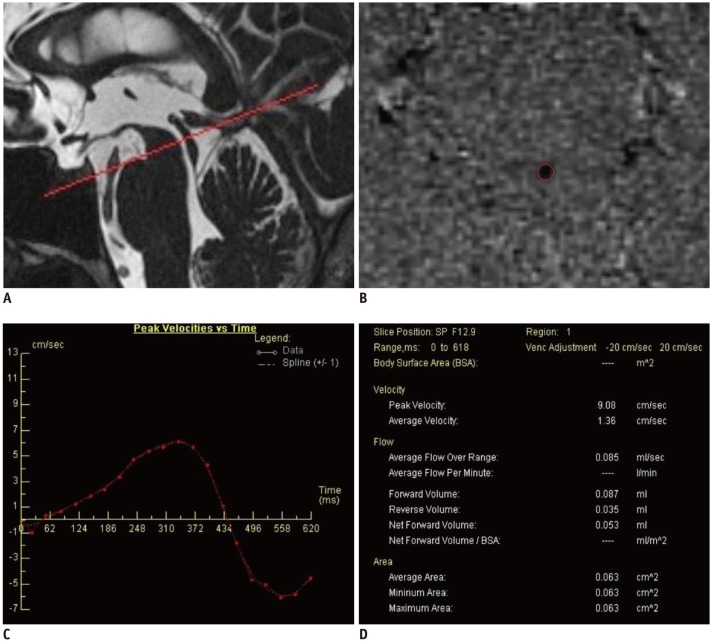
Fig. 1 Measurement of flow of cerebrospinal fluid with phase-contrast MRI.A. Saggital T2 scan showing positioning for cine MRI acquisition. B. Cerebral aqueduct region-of-interest drawing on phase image, after sufficient magnification. C. Peak velocity-time graph obtained for cardiac beat. D. Data table obtained using Argus software.
Reference
-
1. Noseworthy JH, Lucchinetti C, Rodriguez M, Weinshenker BG. Multiple sclerosis. N Engl J Med. 2000; 343:938–952. PMID: 11006371.
Article2. Frohman EM, Racke MK, Raine CS. Multiple sclerosis--the plaque and its pathogenesis. N Engl J Med. 2006; 354:942–955. PMID: 16510748.3. Ge Y, Zohrabian VM, Grossman RI. Seven-Tesla magnetic resonance imaging: new vision of microvascular abnormalities in multiple sclerosis. Arch Neurol. 2008; 65:812–816. PMID: 18541803.4. Kermode AG, Thompson AJ, Tofts P, MacManus DG, Kendall BE, Kingsley DP, et al. Breakdown of the blood-brain barrier precedes symptoms and other MRI signs of new lesions in multiple sclerosis. Pathogenetic and clinical implications. Brain. 1990; 113(Pt 5):1477–1489. PMID: 2245307.5. Kidd D, Barkhof F, McConnell R, Algra PR, Allen IV, Revesz T. Cortical lesions in multiple sclerosis. Brain. 1999; 122(Pt 1):17–26. PMID: 10050891.
Article6. Tan IL, van Schijndel RA, Pouwels PJ, van Walderveen MA, Reichenbach JR, Manoliu RA, et al. MR venography of multiple sclerosis. AJNR Am J Neuroradiol. 2000; 21:1039–1042. PMID: 10871010.7. Fog T. The topography of plaques in multiple sclerosis with special reference to cerebral plaques. Acta Neurol Scand Suppl. 1965; 15:1–161. PMID: 5213727.8. Singh AV, Zamboni P. Anomalous venous blood flow and iron deposition in multiple sclerosis. J Cereb Blood Flow Metab. 2009; 29:1867–1878. PMID: 19724286.
Article9. Zamboni P, Menegatti E, Weinstock-Guttman B, Schirda C, Cox JL, Malagoni AM, et al. CSF dynamics and brain volume in multiple sclerosis are associated with extracranial venous flow anomalies: a pilot study. Int Angiol. 2010; 29:140–148. PMID: 20351670.10. Zamboni P, Galeotti R, Menegatti E, Malagoni AM, Tacconi G, Dall'Ara S, et al. Chronic cerebrospinal venous insufficiency in patients with multiple sclerosis. J Neurol Neurosurg Psychiatry. 2009; 80:392–399. PMID: 19060024.
Article11. Zamboni P, Menegatti E, Weinstock-Guttman B, Schirda C, Cox JL, Malagoni AM, et al. The severity of chronic cerebrospinal venous insufficiency in patients with multiple sclerosis is related to altered cerebrospinal fluid dynamics. Funct Neurol. 2009; 24:133–138. PMID: 20018140.12. Barkhof F, Kouwenhoven M, Scheltens P, Sprenger M, Algra P, Valk J. Phase-contrast cine MR imaging of normal aqueductal CSF flow. Effect of aging and relation to CSF void on modulus MR. Acta Radiol. 1994; 35:123–130. PMID: 8172735.13. Gideon P, Thomsen C, Ståhlberg F, Henriksen O. Cerebrospinal fluid production and dynamics in normal aging: a MRI phase-mapping study. Acta Neurol Scand. 1994; 89:362–366. PMID: 8085434.
Article14. Unal O, Kartum A, Avcu S, Etlik O, Arslan H, Bora A. Cine phase-contrast MRI evaluation of normal aqueductal cerebrospinal fluid flow according to sex and age. Diagn Interv Radiol. 2009; 15:227–231. PMID: 19862673.15. Polman CH, Reingold SC, Edan G, Filippi M, Hartung HP, Kappos L, et al. Diagnostic criteria for multiple sclerosis: 2005 revisions to the “McDonald Criteria”. Ann Neurol. 2005; 58:840–846. PMID: 16283615.
Article16. Kurtzke JF. Rating neurologic impairment in multiple sclerosis: an expanded disability status scale (EDSS). Neurology. 1983; 33:1444–1452. PMID: 6685237.
Article17. Zamboni P, Menegatti E, Bartolomei I, Galeotti R, Malagoni AM, Tacconi G, et al. Intracranial venous haemodynamics in multiple sclerosis. Curr Neurovasc Res. 2007; 4:252–258. PMID: 18045150.
Article18. Ghezzi A, Comi G, Federico A. Chronic cerebro-spinal venous insufficiency (CCSVI) and multiple sclerosis. Neurol Sci. 2011; 32:17–21. PMID: 21161309.
Article19. Ursino M, Lodi CA. A simple mathematical model of the interaction between intracranial pressure and cerebral hemodynamics. J Appl Physiol (1985). 1997; 82:1256–1269. PMID: 9104864.20. Kim J, Thacker NA, Bromiley PA, Jackson A. Prediction of the jugular venous waveform using a model of CSF dynamics. AJNR Am J Neuroradiol. 2007; 28:983–989. PMID: 17494684.21. Schaller B. Physiology of cerebral venous blood flow: from experimental data in animals to normal function in humans. Brain Res Brain Res Rev. 2004; 46:243–260. PMID: 15571768.
Article22. Evans AJ, Iwai F, Grist TA, Sostman HD, Hedlund LW, Spritzer CE, et al. Magnetic resonance imaging of blood flow with a phase subtraction technique. In vitro and in vivo validation. Invest Radiol. 1993; 28:109–115. PMID: 8444566.23. Pelc LR, Pelc NJ, Rayhill SC, Castro LJ, Glover GH, Herfkens RJ, et al. Arterial and venous blood flow: noninvasive quantitation with MR imaging. Radiology. 1992; 185:809–812. PMID: 1438767.
Article24. Baracchini C, Perini P, Calabrese M, Causin F, Rinaldi F, Gallo P. No evidence of chronic cerebrospinal venous insufficiency at multiple sclerosis onset. Ann Neurol. 2011; 69:90–99. PMID: 21280079.
Article25. Simka M, Ludyga T, Kazibudzki M, Latacz P, Swierad M. Multiple sclerosis, an unlikely cause of chronic cerebrospinal venous insufficiency: retrospective analysis of catheter venography. JRSM Short Rep. 2012; 3:56. PMID: 23301144.
Article26. Patti F, Nicoletti A, Leone C, Messina S, D'Amico E, Lo Fermo S, et al. Multiple sclerosis and CCSVI: a population-based case control study. PLoS One. 2012; 7:e41227. PMID: 22870210.
Article27. Sundström P, Wåhlin A, Ambarki K, Birgander R, Eklund A, Malm J. Venous and cerebrospinal fluid flow in multiple sclerosis: a case-control study. Ann Neurol. 2010; 68:255–259. PMID: 20695018.
Article28. Gorucu Y, Albayram S, Balci B, Hasiloglu ZI, Yenigul K, Yargic F, et al. Cerebrospinal fluid flow dynamics in patients with multiple sclerosis: a phase contrast magnetic resonance study. Funct Neurol. 2011; 26:215–222. PMID: 22364942.29. Chiang WW, Takoudis CG, Lee SH, Weis-McNulty A, Glick R, Alperin N. Relationship between ventricular morphology and aqueductal cerebrospinal fluid flow in healthy and communicating hydrocephalus. Invest Radiol. 2009; 44:192–199. PMID: 19300098.
Article30. Nitz WR, Bradley WG Jr, Watanabe AS, Lee RR, Burgoyne B, O'Sullivan RM, et al. Flow dynamics of cerebrospinal fluid: assessment with phase-contrast velocity MR imaging performed with retrospective cardiac gating. Radiology. 1992; 183:395–405. PMID: 1561340.
Article
- Full Text Links
-
- Actions
-
Cited
- CITED
-
- Close
- Share
-
- Similar articles
-
- Treatment of Syringomyelia with Consideration on its Pathophysiology
- Chronic Cerebrospinal Venous Insufficiency in Multiple Sclerosis: A Failed Concept
- Oligoclonal Bands in Cerebrospinal fluid of Neurologic patients
- Understanding MRI Features of Multiple Sclerosis: Based on the 2017 McDonald Criteria
- Assessment of Flow Dynamics of Cerebrospinal Fluid with Phase-contrast Cine MR Image
