Three-Dimensional Fast Spin-Echo Imaging without Fat Suppression of the Knee: Diagnostic Accuracy Comparison to Fat-Suppressed Imaging on 1.5T MRI
- Affiliations
-
- 1Department of Radiology, Research Institute of Radiological Science, YUHS-KRIBB Medical Convergence Research Institute, and Severance Biomedical Science Institute, Yonsei University College of Medicine, Seoul, Korea. jss@yuhs.ac
- 2Department of Orthopedic Surgery, Yonsei University College of Medicine, Seoul, Korea.
- KMID: 2418901
- DOI: http://doi.org/10.3349/ymj.2017.58.6.1186
Abstract
- PURPOSE
To evaluate the diagnostic performance of three-dimensional fast spin-echo (3D FSE-Cube) without fat suppression (NFS) for detecting knee lesions, using comparison to 3D FSE-Cube with fat suppression (FS).
MATERIALS AND METHODS
One hundred twenty-four patients who underwent 1.5T knee magnetic resonance imaging (MRI) scans and 25 subsequent arthroscopic surgeries were retrospectively reviewed. Using arthroscopic results and two-dimensional images as reference standards, diagnostic performances of 3D FSE-Cube-NFS and FS imaging about lesions of ligament, meniscus, subchondral bone marrow edema (BME), and cartilage were compared. Scan parameters of 3D FSE-Cube imaging were previously optimized by a porcine knee phantom.
RESULTS
No significant differences were observed between detection rates of NFS and FS imaging for detecting lesions of meniscus and cartilage (p>0.05). However, NFS imaging had lower sensitivity for detection of medial collateral ligament (MCL) tears, and lower sensitivity and specificity for detection of BME lesions, compared to FS imaging (p<0.05).
CONCLUSION
3D FSE-Cube-NFS imaging showed similar diagnostic performance for detecting lesions of meniscus or cartilage compared to FS imaging, unlike MCL or BME lesions.
MeSH Terms
Figure
-
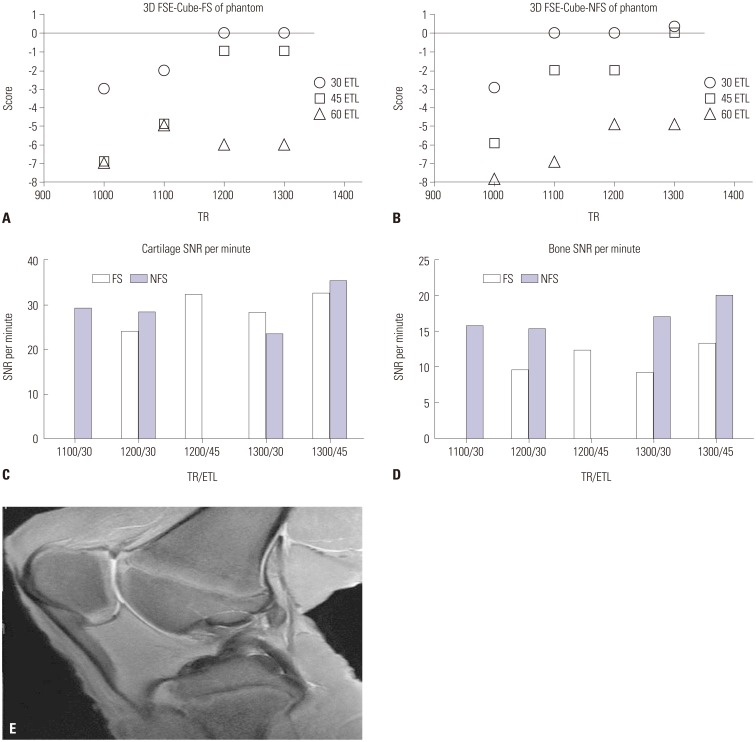
Fig. 1 Subjective scores of phantom imaging (A) with fat suppression and (B) without fat suppression. Images with a score of -1 or above were regarded as acceptable. Highest SNRs per unit time were acquired with scan parameters of TR=1300 ms and ETL=45 in both FS and NFS images with parameter settings of acceptable image quality, measured in the (C) patellar cartilage and (D) femoral epiphyseal bone marrow. (E) 3D FSE-Cube-NFS phantom image with optimized parameters. 3D FSE-Cube-FS, three-dimensional fast spin-echo with fat suppression; 3D FSE-Cube-NFS, three-dimensional fast spin-echo without fat suppression; SNR, signal-to-noise ratio; TR, repetition time; ETL, echo train length.
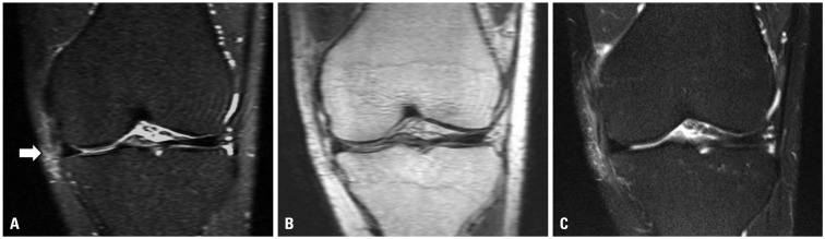
Fig. 2 (A) Blurring of MCL fibers in addition to an increase in signal intensity of surrounding soft tissue are seen on the coronal reformatted 3D FSE-Cube-FS image (arrow). (B) Blurring of ligament fiber is not definite, and no signal abnormality in surrounding soft tissue is detected on the coronal reformatted 3D FSE-Cube-NFS image. (C) Conventional 2D coronal T2-weighted image reveals MCL tear. MCL, medial collateral ligament; 3D FSE-Cube-FS, three-dimensional fast spin-echo with fat suppression; 3D FSE-Cube-NFS, three-dimensional fast spin-echo without fat suppression.
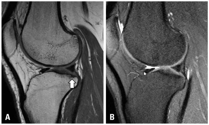
Fig. 3 (A) 3D FSE-Cube-NFS image shows a suspicious subchondral BME lesion on the posterolateral tibial plateau (arrow), while (B) 3D FSE-Cube-FS image shows a subchondral sclerotic change. After correlation with 2D imaging, it turned out to be a false-positive lesion. 3D FSE-Cube-FS, three-dimensional fast spin-echo with fat suppression; 3D FSE-Cube-NFS, three-dimensional fast spin-echo without fat suppression; 2D, two-dimensional.
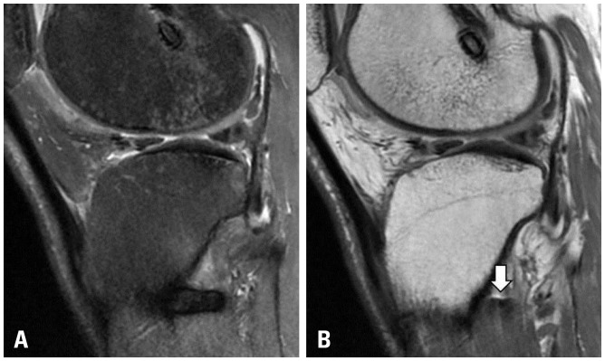
Fig. 4 (A) Metal artifact posterior to the proximal tibial cortex in 3D FSE-Cube-FS image. (B) Greatly reduced artifact is noted in 3D FSE-Cube-NFS image (arrow). Posterior tibial cortex margin is well-demarcated in this sequence. 3D FSE-Cube-FS, three-dimensional fast spin-echo with fat suppression; 3D FSE-Cube-NFS, three-dimensional fast spin-echo without fat suppression.
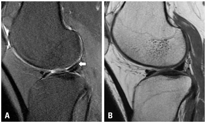
Fig. 5 (A) Artifact during 3D FSE-Cube-FS imaging. It is impossible to know whether there is a tear in the LM (arrow). (B) Subsequent 3D FSE-Cube-NFS image shows a normal meniscus. 3D FSE-Cube-FS, three-dimensional fast spin-echo with fat suppression; 3D FSE-Cube-NFS, three-dimensional fast spin-echo without fat suppression; LM, lateral meniscus.
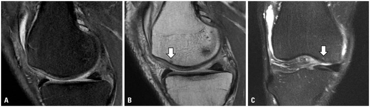
Fig. 6 (A) No definite subchondral bone marrow signal change is seen in the medial femoral condyle in 3D FSE-Cube-FS image. (B) A small suspicious low signal intensity lesion is seen in the subsequent 3D FSE-Cube-NFS image (arrow). (C) Conventional 2D coronal T2-weighted image reveals a tiny subchondral BME lesion in the medial femoral condyle (arrow). 3D FSE-Cube-FS, three-dimensional fast spin-echo with fat suppression; 3D FSE-Cube-NFS, three-dimensional fast spin-echo without fat suppression; 2D, two-dimensional.
Reference
-
1. Mackenzie R, Dixon AK, Keene GS, Hollingworth W, Lomas DJ, Villar RN. Magnetic resonance imaging of the knee: assessment of effectiveness. Clin Radiol. 1996; 51:245–250. PMID: 8617035.
Article2. Gold GE, Busse RF, Beehler C, Han E, Brau AC, Beatty PJ, et al. Isotropic MRI of the knee with 3D fast spin-echo extended echo-train acquisition (XETA): initial experience. AJR Am J Roentgenol. 2007; 188:1287–1293. PMID: 17449772.
Article3. Kijowski R, Blankenbaker DG, Woods M, Del Rio AM, De Smet AA, Reeder SB. Clinical usefulness of adding 3D cartilage imaging sequences to a routine knee MR protocol. AJR Am J Roentgenol. 2011; 196:159–167. PMID: 21178062.
Article4. Kijowski R, Davis KW, Woods MA, Lindstrom MJ, De Smet AA, Gold GE, et al. Knee joint: comprehensive assessment with 3D isotropic resolution fast spin-echo MR imaging--diagnostic performance compared with that of conventional MR imaging at 3.0 T. Radiology. 2009; 252:486–495. PMID: 19703886.
Article5. Jung JY, Jee WH, Park MY, Lee SY, Kim JM. Meniscal tear configurations: categorization with 3D isotropic turbo spin-echo MRI compared with conventional MRI at 3 T. AJR Am J Roentgenol. 2012; 198:W173–W180. PMID: 22268208.
Article6. Jung JY, Yoon YC, Kim HR, Choe BK, Wang JH, Jung JY. Knee derangements: comparison of isotropic 3D fast spin-echo, isotropic 3D balanced fast field-echo, and conventional 2D fast spin-echo MR imaging. Radiology. 2013; 268:802–813. PMID: 23533289.
Article7. Jung JY, Yoon YC, Kwon JW, Ahn JH, Choe BK. Diagnosis of internal derangement of the knee at 3.0-T MR imaging: 3D isotropic intermediate-weighted versus 2D sequences. Radiology. 2009; 253:780–787. PMID: 19789228.
Article8. Lim D, Han Lee Y, Kim S, Song HT, Suh JS. Clinical value of fat-suppressed 3D volume isotropic spin-echo (VISTA) sequence compared to 2D sequence in evaluating internal structures of the knee. Acta Radiol. 2016; 57:66–73. PMID: 25585850.
Article9. Ai T, Zhang W, Priddy NK, Li X. Diagnostic performance of CUBE MRI sequences of the knee compared with conventional MRI. Clin Radiol. 2012; 67:e58–e63. PMID: 22974569.
Article10. Kijowski R, Davis KW, Blankenbaker DG, Woods MA, Del Rio AM, De Smet AA. Evaluation of the menisci of the knee joint using three-dimensional isotropic resolution fast spin-echo imaging: diagnostic performance in 250 patients with surgical correlation. Skeletal Radiol. 2012; 41:169–178. PMID: 21399933.
Article11. Ristow O, Steinbach L, Sabo G, Krug R, Huber M, Rauscher I, et al. Isotropic 3D fast spin-echo imaging versus standard 2D imaging at 3.0 T of the knee--image quality and diagnostic performance. Eur Radiol. 2009; 19:1263–1272. PMID: 19137309.
Article12. Duc SR, Pfirrmann CW, Koch PP, Zanetti M, Hodler J. Internal knee derangement assessed with 3-minute three-dimensional isovoxel true FISP MR sequence: preliminary study. Radiology. 2008; 246:526–535. PMID: 18227545.
Article13. Lim D, Lee YH, Kim S, Song HT, Suh JS. Fat-suppressed volume isotropic turbo spin echo acquisition (VISTA) MR imaging in evaluating radial and root tears of the meniscus: focusing on reader-defined axial reconstruction. Eur J Radiol. 2013; 82:2296–2302. PMID: 24074646.
Article14. Delfaut EM, Beltran J, Johnson G, Rousseau J, Marchandise X, Cotten A. Fat suppression in MR imaging: techniques and pitfalls. Radiographics. 1999; 19:373–382. PMID: 10194785.
Article15. Cho KE, Yoon CS, Song HT, Lee YH, Lim D, Suh JS, et al. Quantitative assessment and ligament traceability of volume isotropic turbo spin echo acquisition (VISTA) ankle magnetic resonance imaging: fat suppression versus without fat suppression. J Korean Soc Magn Reson Med. 2013; 17:110–122.
Article16. Schäfer FK, Schäfer PJ, Brossmann J, Frahm C, Hilgert RE, Heller M, et al. Value of fat-suppressed proton-density-weighted turbo spin-echo sequences in detecting meniscal lesions: comparison with arthroscopy. Acta Radiol. 2006; 47:385–390. PMID: 16739698.
Article17. Lee JH, Yoon YC, Park KJ, Wang JH. Diagnosis of internal derangement of the knee: volume isotropic turbo spin-echo acquisition MRI with fat suppression versus without fat suppression. AJR Am J Roentgenol. 2017; 208:1304–1311. PMID: 28301221.
Article18. Lee SY, Jee WH, Kim SK, Kim JM. Proton density-weighted MR imaging of the knee: fat suppression versus without fat suppression. Skeletal Radiol. 2011; 40:189–195. PMID: 20512570.
Article19. Li CQ, Chen W, Rosenberg JK, Beatty PJ, Kijowski R, Hargreaves BA, et al. Optimizing isotropic three-dimensional fast spin-echo methods for imaging the knee. J Magn Reson Imaging. 2014; 39:1417–1425. PMID: 24987753.
Article20. De Smet AA, Norris MA, Yandow DR, Quintana FA, Graf BK, Keene JS. MR diagnosis of meniscal tears of the knee: importance of high signal in the meniscus that extends to the surface. AJR Am J Roentgenol. 1993; 161:101–107. PMID: 8517286.
Article21. De Smet AA, Tuite MJ, Norris MA, Swan JS. MR diagnosis of meniscal tears: analysis of causes of errors. AJR Am J Roentgenol. 1994; 163:1419–1423. PMID: 7992739.
Article22. Bredella MA, Tirman PF, Peterfy CG, Zarlingo M, Feller JF, Bost FW, et al. Accuracy of T2-weighted fast spin-echo MR imaging with fat saturation in detecting cartilage defects in the knee: comparison with arthroscopy in 130 patients. AJR Am J Roentgenol. 1999; 172:1073–1080. PMID: 10587150.
Article23. Potter HG, Linklater JM, Allen AA, Hannafin JA, Haas SB. Magnetic resonance imaging of articular cartilage in the knee. An evaluation with use of fast-spin-echo imaging. J Bone Joint Surg Am. 1998; 80:1276–1284. PMID: 9759811.
Article24. Sonin AH, Pensy RA, Mulligan ME, Hatem S. Grading articular cartilage of the knee using fast spin-echo proton density-weighted MR imaging without fat suppression. AJR Am J Roentgenol. 2002; 179:1159–1166. PMID: 12388492.
Article
- Full Text Links
-
- Actions
-
Cited
- CITED
-
- Close
- Share
-
- Similar articles
-
- Meniscal Tears of the Knee: Diagnosis with Fast Spin-Echo MR Imaging and Role of Gadolinium-Enhancement
- Diagnosis of Meniscal Tear of the Knee Using Proton-weighted Fast Spin-Echo MR Imaging: Can be an Alternative to Conventional Spin-Echo Imaging?
- Evaluation of the Chondromalacia Patella Using a Microscopy Coil: Comparison of the Two-Dimensional Fast Spin Echo Techniques and the Three-Dimensional Fast Field Echo Techniques
- Fast Spin-Echo T2-Weighted MR Imaging of Tongue Cancer: the Value of Fat-suppression
- MR Imaging of the Spine at 3.0T with T2-Weighted IDEAL Fast Recovery Fast Spin-Echo Technique
