MR Imaging of the Spine at 3.0T with T2-Weighted IDEAL Fast Recovery Fast Spin-Echo Technique
- Affiliations
-
- 1Department of Radiology, Navy General Hospital of PLA, Beijing 100048, Republic of China. nrren@yahoo.com
- KMID: 1058792
- DOI: http://doi.org/10.3348/kjr.2012.13.1.44
Abstract
OBJECTIVE
To compare the iterative decomposition of water and fat with echo asymmetry and the least-squares estimation (IDEAL) method with a fat-saturated T2-weighted (T2W) fast recovery fast spin-echo (FRFSE) imaging of the spine.
MATERIALS AND METHODS
Images acquired at 3.0 Tesla (T) in 35 patients with different spine lesions using fat-saturated T2W FRFSE imaging were compared with T2W IDEAL FRFSE images. Signal-to-noise ratio (SNR)-efficiencies measurements were made in the vertebral bodies and spinal cord in the mid-sagittal plane or nearest to the mid-sagittal plane. Images were scored with the consensus of two experienced radiologists on a four-point grading scale for fat suppression and overall image quality. Statistical analysis of SNR-efficiency, fat suppression and image quality scores was performed with a paired Student's t test and Wilcoxon's signed rank test.
RESULTS
Signal-to-noise ratio-efficiency for both vertebral body and spinal cord was higher with T2W IDEAL FRFSE imaging (p < 0.05) than with T2W FRFSE imaging. T2W IDEAL FRFSE demonstrated superior fat suppression (p < 0.01) and image quality (p < 0.01) compared to fat-saturated T2W FRFSE.
CONCLUSION
As compared with fat-saturated T2W FRFSE, IDEAL can provide a higher image quality, higher SNR-efficiency, and consistent, robust and uniform fat suppression. T2W IDEAL FRFSE is a promising technique for MR imaging of the spine at 3.0T.
Keyword
MeSH Terms
Figure
-

Fig. 1 41-year-old woman with lower back pain. A. Sagittal T2-weighted iterative decomposition of water and fat with echo asymmetry and least-squares estimation (IDEAL) fast recovery fast spin-echo (FRFSE) water-only image. B. Iterative decomposition of water and fat with echo asymmetry and least-squares estimation (IDEAL) fat-only image. C. Recombined iterative decomposition of water and fat with echo asymmetry and least-squares estimation (IDEAL) in-phase image. D. Recombined iterative decomposition of water and fat with echo asymmetry and least-squares estimation (IDEAL) out-of-phase image. Separate water (A) and fat (B) images demonstrate uniform separation of water and fat. Correction of chemical shift artifacts is shown in recombined in-phase image (C).

Fig. 2 26-year-old man with history of surgery after traffic accident. A. Sagittal T2-weighted iterative decomposition of water and fat with echo asymmetry and least-squares estimation (IDEAL) fast recovery fast spin-echo (FRFSE) water-only image. B. Sagittal fat-saturated T2-weighted fast recovery fast spin-echo (FRFSE) image. Failed fat-saturation and susceptibility artifacts due to B0 field inhomogeneities generated from metallic hardware in several thoracic vertebral bodies that obscured large portion of spinal cord and vertebral body (arrows). Iterative decomposition of water and fat with echo asymmetry and least-squares estimation (IDEAL) image demonstrated very uniform fat separation despite presence of metallic hardware (A).
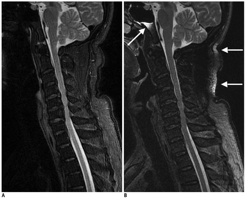
Fig. 3 56-year-old man with neck pain. A. Sagittal T2-weighted iterative decomposition of water and fat with echo asymmetry and least-squares estimation (IDEAL) fast recovery fast spin-echo (FRFSE) water-only image. B. Sagittal fat-saturated T2-weighted fast recovery fast spin-echo (FRFSE) image. Note failure of fat suppression in areas with air-tissue interfaces, and unfavorable geometry on fat-saturated fast recovery fast spin-echo (FRFSE) image (arrows), but uniformity of water-fat separation on water-only image was demonstrated (A).
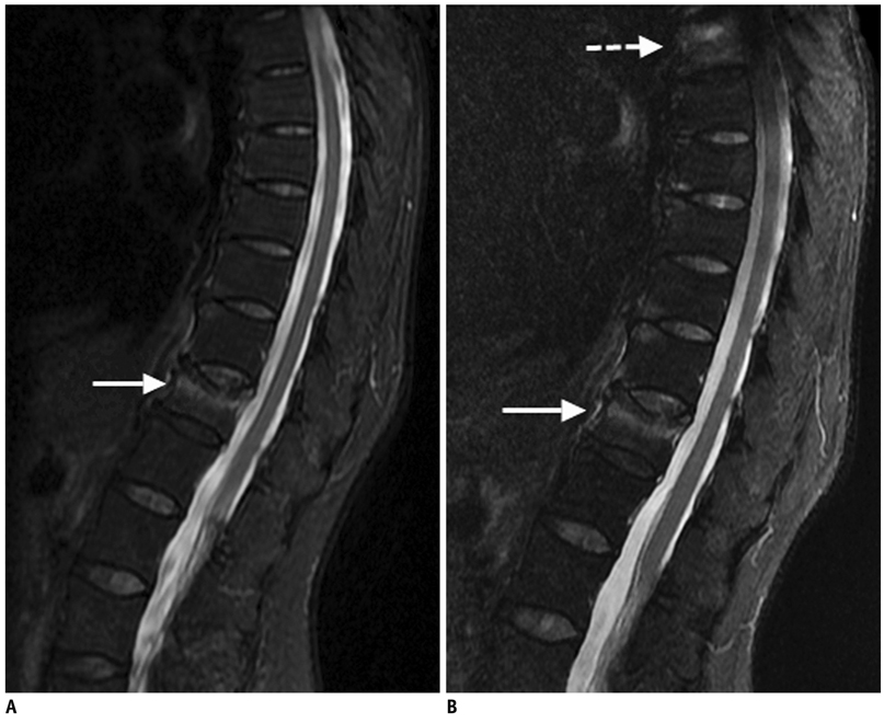
Fig. 4 76-year-old woman with history of trauma. A. Sagittal T2-weighted iterative decomposition of water and fat with echo asymmetry and least-squares estimation (IDEAL) fast recovery fast spin-echo (FRFSE) water-only image. B. Sagittal fat-saturated T2-weighted fast recovery fast spin-echo (FRFSE) image. Compression of T11 vertebral body is seen (arrows). Poor fat suppression on fat-saturated fast recovery fast spin-echo (FRFSE) image in areas near top margin of field of view is seen mimicking T5 vertebral body marrow edema (dashed arrow), but superior fat-water separation and signal-to-noise ratio performance in corresponding iterative decomposition of water and fat with echo asymmetry and least-squares estimation (IDEAL) water-only image show that this is artifact caused by poor fat saturation (A).

Fig. 5 56-year-old man with 12-year history of lung adenocarcinoma. A, B. Sagittal T2-weighted iterative decomposition of water and fat with echo asymmetry and least-squares estimation (IDEAL) fast recovery fast spin-echo (FRFSE) water-only images. C, D. Corresponding fat-saturated T2-weighted fast recovery fast spin-echo (FRFSE) images. This patient has multiple metastases in spine. Fat-saturated T2-weighted fast recovery fast spin-echo (FRFSE) images demonstrate unreliable fat suppression in superior regions of images due to off-isocenter B0 inhomogeneities (arrows), which obscured lesion in left spinal facet joint at T9/T10 (dashed arrow). All lesions are distinctly visualized on iterative decomposition of water and fat with echo asymmetry and least-squares estimation (IDEAL) water-only images because of uniform and reliable fat suppression (arrowheads).
Cited by 1 articles
-
In Vivo Assessment of Neurodegeneration in Type C Niemann-Pick Disease by IDEAL-IQ
Ruo-Mi Guo, Qing-Ling Li, Zhong-Xing Luo, Wen Tang, Ju Jiao, Jin Wang, Zhuang Kang, Shao-Qiong Chen, Yong Zhang
Korean J Radiol. 2018;19(1):93-100. doi: 10.3348/kjr.2018.19.1.93.
Reference
-
1. Vertinsky AT, Krasnokutsky MV, Augustin M, Bammer R. Cutting-edge imaging of the spine. Neuroimaging Clin N Am. 2007. 17:117–136.2. Alyas F, Saifuddin A, Connell D. MR imaging evaluation of the bone marrow and marrow infiltrative disorders of the lumbar spine. Magn Reson Imaging Clin N Am. 2007. 15:199–219.3. Nobauer I, Uffmann M. Differential diagnosis of focal and diffuse neoplastic diseases of bone marrow in MRI. Eur J Radiol. 2005. 55:2–32.4. Vanel D, Dromain C, Tardivon A. MRI of bone marrow disorders. Eur Radiol. 2000. 10:224–229.5. D'Aprile P, Tarantino A, Jinkins JR, Brindicci D. The value of fat saturation sequences and contrast medium administration in MRI of degenerative disease of the posterior/perispinal elements of the lumbosacral spine. Eur Radiol. 2007. 17:523–531.6. Reeder SB, Yu H, Johnson JW, Shimakawa A, Brittain JH, Pelc NJ, et al. T1- and T2-weighted fast spin-echo imaging of the brachial plexus and cervical spine with IDEAL water-fat separation. J Magn Reson Imaging. 2006. 24:825–832.7. Zha Y, Li M, Yang J. Dynamic contrast enhanced magnetic resonance imaging of diffuse spinal bone marrow infiltration in patients with hematological malignancies. Korean J Radiol. 2010. 11:187–194.8. Oner AY, Akpek S, Tali T, Ucar M. Giant vertebral notochordal rest: magnetic resonance and diffusion weighted imaging findings. Korean J Radiol. 2009. 10:303–306.9. Reeder SB, Pineda AR, Wen Z, Shimakawa A, Yu H, Brittain JH, et al. Iterative decomposition of water and fat with echo asymmetry and least-squares estimation (IDEAL): application with fast spin-echo imaging. Magn Reson Med. 2005. 54:636–644.10. Bydder GM, Pennock JM, Steiner RE, Khenia S, Payne JA, Young IR. The short TI inversion recovery sequence--an approach to MR imaging of the abdomen. Magn Reson Imaging. 1985. 3:251–254.11. Block W, Pauly J, Kerr A, Nishimura D. Consistent fat suppression with compensated spectral-spatial pulses. Magn Reson Med. 1997. 38:198–206.12. Dixon WT. Simple proton spectroscopic imaging. Radiology. 1984. 153:189–194.13. Glover GH. Multipoint Dixon technique for water and fat proton and susceptibility imaging. J Magn Reson Imaging. 1991. 1:521–530.14. Glover GH, Schneider E. Three-point Dixon technique for true water/fat decomposition with B0 inhomogeneity correction. Magn Reson Med. 1991. 18:371–383.15. Hardy PA, Hinks RS, Tkach JA. Separation of fat and water in fast spin-echo MR imaging with the three-point Dixon technique. J Magn Reson Imaging. 1995. 5:181–185.16. Pineda AR, Reeder SB, Wen Z, Pelc NJ. Cramer-Rao bounds for three-point decomposition of water and fat. Magn Reson Med. 2005. 54:625–635.17. Reeder SB, Wen Z, Yu H, Pineda AR, Gold GE, Markl M, et al. Multicoil Dixon chemical species separation with an iterative least-squares estimation method. Magn Reson Med. 2004. 51:35–45.18. Kijowski R, Blankenbaker DG, Woods MA, Shinki K, De Smet AA, Reeder SB. 3.0-T evaluation of knee cartilage by using three-dimensional IDEAL GRASS imaging: comparison with fast spin-echo imaging. Radiology. 2010. 255:117–127.19. Reeder SB, McKenzie CA, Pineda AR, Yu H, Shimakawa A, Brau AC, et al. Water-fat separation with IDEAL gradient-echo imaging. J Magn Reson Imaging. 2007. 25:644–652.20. Gold GE, Reeder SB, Yu H, Kornaat P, Shimakawa AS, Johnson JW, et al. Articular cartilage of the knee: rapid three-dimensional MR imaging at 3.0 T with IDEAL balanced steady-state free precession--initial experience. Radiology. 2006. 240:546–551.21. Barger AV, DeLone DR, Bernstein MA, Welker KM. Fat signal suppression in head and neck imaging using fast spin-echo-IDEAL technique. AJNR Am J Neuroradiol. 2006. 27:1292–1294.22. Yu H, Reeder SB, McKenzie CA, Brau AC, Shimakawa A, Brittain JH, et al. Single acquisition water-fat separation: feasibility study for dynamic imaging. Magn Reson Med. 2006. 55:413–422.23. Reeder SB, Markl M, Yu H, Hellinger JC, Herfkens RJ, Pelc NJ. Cardiac CINE imaging with IDEAL water-fat separation and steady-state free precession. J Magn Reson Imaging. 2005. 22:44–52.24. Fuller S, Reeder S, Shimakawa A, Yu H, Johnson J, Beaulieu C, et al. Iterative decomposition of water and fat with echo asymmetry and least-squares estimation (IDEAL) fast spine-cho imaging of the ankle: initial clinical experience. AJR Am J Roentgenol. 2006. 187:1442–1447.25. Murakami M, Mori H, Kunimatsu A, Abe O, Chikuda H, Ono T, et al. Postsurgical spinal magnetic resonance imaging with iterative decomposition of water and fat with echo asymmetry and least-squares estimation. J Comput Assist Tomogr. 2011. 35:16–20.26. Zajick DC Jr, Morrison WB, Schweitzer ME, Parellada JA, Carrino JA. Benign and malignant processes: normal values and differentiation with chemical shift MR imaging in vertebral marrow. Radiology. 2005. 237:590–596.
- Full Text Links
-
- Actions
-
Cited
- CITED
-
- Close
- Share
-
- Similar articles
-
- Diagnosis of Meniscal Tear of the Knee Using Proton-weighted Fast Spin-Echo MR Imaging: Can be an Alternative to Conventional Spin-Echo Imaging?
- Meniscal Tears of the Knee: Diagnosis with Fast Spin-Echo MR Imaging and Role of Gadolinium-Enhancement
- Fast FLAIR MR Imaging Finidngs of Cerebral Infarction: Comparison with T2-Weighted Spin Echo Imaging
- The evaluation of Fat Saturation Fast Spin-Echo T2WI for Patients with Acute Spinal Trauma
- A study of artifacts in MR imaging induced by metalic aneurysm clips
