Yonsei Med J.
2018 Mar;59(2):252-257. 10.3349/ymj.2018.59.2.252.
Coronary Flow Reserve in Non-Infarcted Myocardium Predicts Long-Term Clinical Outcomes in Patients Undergoing Percutaneous Coronary Intervention
- Affiliations
-
- 1Department of Cardiology, the Forth Affiliated Hospital of Harbin Medical University, Harbin, China. chengrongchao@163.com
- 2Department of Economic Management, Heilongjiang Nongken Vocational College, Harbin, China.
- KMID: 2418788
- DOI: http://doi.org/10.3349/ymj.2018.59.2.252
Abstract
- PURPOSE
Coronary flow reserve (CFR) is recognized as an indicator of myocardial perfusion. The aim of this study was to assess the relationship between CFR in the non-infarcted myocardium and the incidence of major adverse cardiac events (MACEs).
MATERIALS AND METHODS
100 consecutive patients with acute myocardial infarction (AMI) undergoing percutaneous coronary intervention (PCI) were enrolled in the present study, and divided into MACE and non-MACE groups according to the incidence of 12-month MACEs. Left ventricular function and CFR were analyzed using two-dimensional echocardiography and myocardial contrast echocardiography at one week after PCI. Cardiac troponin I levels were assayed to estimate peak concentrations thereof.
RESULTS
The MACE group was associated with lower CFR, compared to the non-MACE group (2.41 vs. 2.77, p < 0.001). In the multivariable model, CFR in the non-infarcted myocardium was an independent predictor of 12-month MACE (hazard ratio: 0.093, 95% confidence interval: 0.020-0.426, p=0.002) after adjustment for baseline demographic and clinical characteristics.
CONCLUSION
CFR in the non-infarcted myocardium is a useful marker for predicting 12-month MACEs in patients with AMI undergoing primary PCI.
Keyword
MeSH Terms
-
Aged
Coronary Circulation/*physiology
*Echocardiography
Female
Fractional Flow Reserve, Myocardial
Humans
Male
Middle Aged
Myocardial Infarction/diagnostic imaging/*physiopathology/*surgery
Myocardial Perfusion Imaging
Myocardium/*pathology
*Percutaneous Coronary Intervention
Proportional Hazards Models
Treatment Outcome
Ventricular Function, Left/*physiology
Figure
-

Fig. 1 The best cut-off values of CFR in the non-infarcted myocardium and peak cTnI were analyzed using ROC curves. CFR in the non-infarcted region (AUC=0.958), cut-off=2.305, sensitivity=0.91, specificity=0.86). Peak cTnI (AUC=0.935), cut-off=18.50, senstivity=0.80, specificity=0.94. CFR, coronary flow reserve; cTnI, cardiac troponin I; ROC, receiver-operating characteristic; AUC, area under the curve.
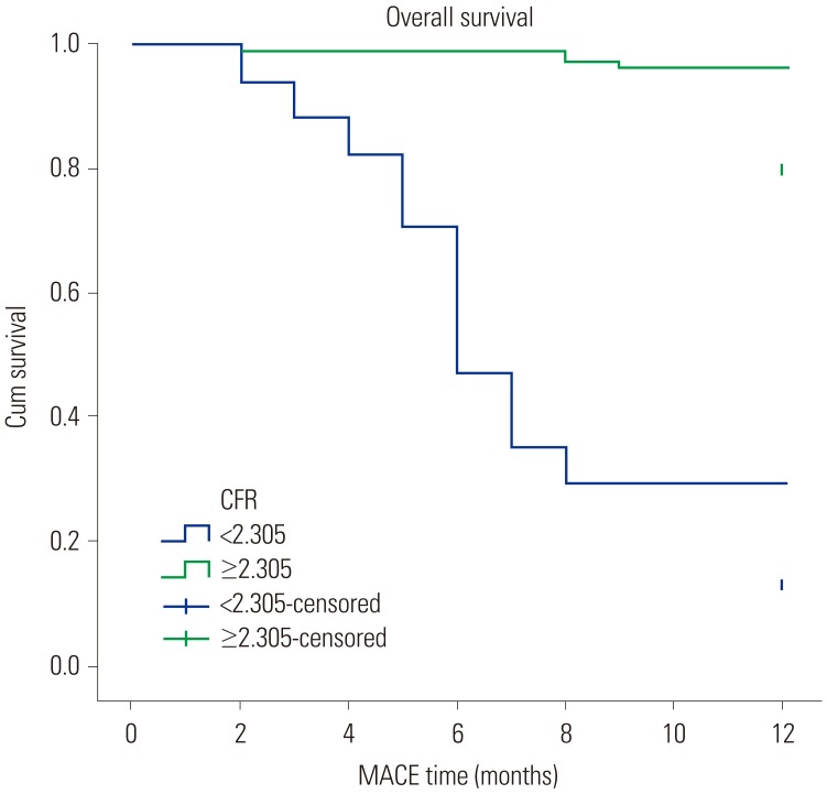
Fig. 2 Kaplan-Meier curves of cumulative survival (one-year survival). The results showed median survival times of 7.353±0.802 months in the CFR <2.305 group and 11.795±0.133 months in the CFR ≥2.305 group (p<0.001). MACE, major adverse cardiac event; CFR, coronary flow reserve.
Reference
-
1. Bowers TR, O'Neill WW. Coronary rotablation and reserve: can they occur together. Cathet Cardiovasc Diagn. 1995; 36:277. PMID: 8542642.
Article2. Bierig SM, Mikolajczak P, Herrmann SC, Elmore N, Kern M, Labovitz AJ. Comparison of myocardial contrast echocardiography derived myocardial perfusion reserve with invasive determination of coronary flow reserve. Eur J Echocardiogr. 2009; 10:250–255. PMID: 18723849.
Article3. Kisanuki A, Yuasa T, Kuwahara E, Takasaki K, Yoshifuku S, Otsuji Y, et al. Reproducibility of intravenous intermittent triggered myocardial contrast echocardiography in healthy subjects. Jpn Heart J. 2004; 45:461–473. PMID: 15240966.
Article4. Kaul S. Myocardial contrast echocardiography: basic principles. Prog Cardiovasc Dis. 2001; 44:1–11. PMID: 11533923.
Article5. Wita K, Filipecki A, Lelek M, Bochenek T, Elžbieciak M, Wróbel W, et al. Prediction of left ventricular remodeling in patients with STEMI treated with primary PCI: use of quantitative myocardial contrast echocardiography. Coron Artery Dis. 2011; 22:171–178. PMID: 21394026.6. Porter TR, D’Sa A, Turner C, Jones LA, Minisi AJ, Mohanty PK, et al. Myocardial contrast echocardiography for the assessment of coronary blood flow reserve: validation in humans. J Am Coll Cardiol. 1993; 21:349–355. PMID: 8425997.
Article7. Swinburn JM, Lahiri A, Senior R. Intravenous myocardial contrast echocardiography predicts recovery of dysynergic myocardium early after acute myocardial infarction. J Am Coll Cardiol. 2001; 38:19–25. PMID: 11451273.
Article8. Mengozzi G, Rossini R, Palagi C, Musumeci G, Petronio A, Limbruno U, et al. Usefulness of intravenous myocardial contrast echocardiography in the early left ventricular remodeling in acute myocardial infarction. Am J Cardiol. 2002; 90:713–719. PMID: 12356383.9. Yamamuro A, Akasaka T, Tamita K, Yamabe K, Katayama M, Takagi T, et al. Coronary flow velocity pattern immediately after percutaneous coronary intervention as a predictor of complications and in-hospital survival after acute myocardial infarction. Circulation. 2002; 106:3051–3056. PMID: 12473550.
Article10. Camici PG, Crea F. Coronary microvascular dysfunction. N Engl J Med. 2007; 356:830–840. PMID: 17314342.
Article11. Wu JC, Yun JJ, Dione DP, Heller EN, Deckelbaum LI, Sinusas AJ. Severe regional ischemia alters coronary flow reserve in the remote perfusion area. J Nucl Cardiol. 2000; 7:43–52. PMID: 10698234.
Article12. Kern MJ, Bach RG, Mechem CJ, Caracciolo EA, Aguirre FV, Miller LW, et al. Variations in normal coronary vasodilatory reserve stratified by artery, gender, heart transplantation and coronary artery disease. J Am Coll Cardiol. 1996; 28:1154–1160. PMID: 8890809.
Article13. Bokor D. Diagnostic efficacy of SonoVue. Am J Cardiol. 2000; 86:19–24.
Article14. Eliasen P, Amtorp O. Effect of intracoronary adenosine upon regional blood flow, microvascular blood volume and hematocrit in canine myocardium. Int J Microcirc Clin Exp. 1984; 3:3–12. PMID: 6480228.15. Wei K, Jayaweera AR, Firoozan S, Linka A, Skyba DM, Kaul S. Quantification of myocardial blood flow with ultrasound-induced destruction of microbubbles administered as a constant venous infusion. Circulation. 1998; 97:473–483. PMID: 9490243.
Article16. Tiemann K, Pohl C, Schlosser T, Goenechea J, Bruce M, Veltmann C, et al. Stimulated acoustic emission: pseudo-Doppler shifts seen during the destruction of nonmoving microbubbles. Ultrasound Med Biol. 2000; 26:1161–1167. PMID: 11053751.
Article17. Kaul S. Myocardial contrast echocardiography: 15 years of research and development. Circulation. 1997; 96:3745–3760. PMID: 9396479.18. Galiuto L, Garramone B, Scarà A, Rebuzzi AG, Crea F, La Torre G, et al. The extent of microvascular damage during myocardial contrast echocardiography is superior to other known indexes of post-infarct reperfusion in predicting left ventricular remodeling: results of the multicenter AMICI study. J Am Coll Cardiol. 2008; 51:552–559. PMID: 18237684.19. Yang L, Xia C, Mu Y, Guan L, Wang C, Tang Q, et al. Prognostic value of real time myocardial contrast echocardiography after percutaneous coronary intervention. Echocardiography. 2016; 33:421–430. PMID: 26411872.
Article20. Duffy BK, Gurm HS, Rajagopal V, Gupta R, Ellis SG, Bhatt DL. Usefulness of an elevated neutrophil to lymphocyte ratio in predicting long-term mortality after percutaneous coronary intervention. Am J Cardiol. 2006; 97:993–996. PMID: 16563903.
Article21. Aoki H, Matsunari I, Nomura Y, Fujita W, Komatsu R, Miyazaki Y, et al. Myocardial sympathetic innervation, function, and oxidative metabolism in non-infarcted myocardium in patients with prior myocardial infarction. Ann Nucl Med. 2013; 27:523–531. PMID: 23494212.
Article22. Feldman RL, Macdonald RG, Nichols WW, Conti CR, Pepine CJ. Effects of acute coronary occlusion on hemodynamics in an adjacent coronary artery in dogs. Am J Cardiol. 1984; 54:1103–1107. PMID: 6496331.
Article23. Uren NG, Crake T, Lefroy DC, de Silva R, Davies GJ, Maseri A. Reduced coronary vasodilator function in infarcted and normal myocardium after myocardial infarction. N Engl J Med. 1994; 331:222–227. PMID: 7832835.
Article24. Neizel M, Futterer S, Steen H, Giannitsis E, Reinhardt L, Lossnitzer D, et al. Predicting microvascular obstruction with cardiac troponin T after acute myocardial infarction: a correlative study with contrast-enhanced magnetic resonance imaging. Clin Res Cardiol. 2009; 98:555–562. PMID: 19582365.
Article25. de Boer RA, Pinto YM, Suurmeijer AJ, Pokharel S, Scholtens E, Humler M, et al. Increased expression of cardiac angiotensin II type 1 (AT(1)) receptors decreases myocardial microvessel density after experimental myocardial infarction. Cardiovasc Res. 2003; 57:434–442. PMID: 12566116.26. Tsai CF, Ueng KC, Wu DJ, Tsai TP, Lin CS. Remodeled left ventricular myocardium remote to infarction sites is the arrhythmogenic substrate for sudden cardiac death. Med Hypotheses. 2010; 75:368–371. PMID: 20399566.
Article27. Bekkers SC, Yazdani SK, Virmani R, Waltenberger J. Microvascular obstruction: underlying pathophysiology and clinical diagnosis. J Am Coll Cardiol. 2010; 55:1649–1660. PMID: 20394867.
- Full Text Links
-
- Actions
-
Cited
- CITED
-
- Close
- Share
-
- Similar articles
-
- Percutaneous coronary intervention in patients with multi-vessel coronary artery disease: a focus on physiology
- Coronary Flow Reserve in the Remote Myocardium Predicts Left Ventricular Remodeling Following Acute Myocardial Infarction
- Relation between Perfusion Status of Dysfunctional Myocardium and Coronary Flow Reserve in Acute Myocardial Infarction
- Coronary Flow Velocity Pattern in Patients with Myocardial Bridging of Coronary Artery
- Physiologic Evaluation of Microvascular Damage in Culprit Vessel After Successful Primary Percutaneous Coronary Intervention for ST-elevation Myocardial Infarction Patients
