Korean J Radiol.
2018 Jun;19(3):489-497. 10.3348/kjr.2018.19.3.489.
Two Small Intravenous Catheters for High-Rate Contrast Medium Injection for Computed Tomography in Patients Lacking Superficial Veins to Accommodate a Large Catheter
- Affiliations
-
- 1Department of Radiology and Research Institute of Radiology, University of Ulsan College of Medicine, Asan Medical Center, Seoul 05505, Korea. parksh.radiology@gmail.com
- KMID: 2410820
- DOI: http://doi.org/10.3348/kjr.2018.19.3.489
Abstract
OBJECTIVE
To prospectively investigate the feasibility of using 2 small intravenous catheters for high-rate computed tomography (CT) contrast injection in patients lacking superficial veins capable of accommodating ≤ 20-gauge catheters.
MATERIALS AND METHODS
Sixty-eight consecutive eligible adults referred for dynamic liver CT were enrolled; 58 had previously undergone liver CT, including 8 that experienced extravasation. Two 22- or 24-gauge catheters were placed in all patients after 2-5 venipunctures, and 2 mL/kg of contrast agent (370 mg I/mL) was split-administered through both catheters to achieve total flow rate of 4 mL/s. Patients' experience and examination success rate, defined as uneventful scans completed at 4 mL/s or at < 4 mL/s achieving standard image quality in all phases, were analyzed. Quantitative hepatic signal-to-noise and hepatic vascular contrast-to-noise ratios (CNRs) were compared with 30 control examinations scanned at 4 mL/s using an 18-gauge catheter.
RESULTS
One case each of extravasation and severe injection pain caused the examination to be aborted. Success rate was 88.2% (60/68; 54 patients scanned at 4 mL/s, 6 at 3.5-3.9 mL/s). Fifty-five of 58 patients (94.8%) that had past CT regarded the venipuncture as more tolerable than (n = 36) or similar to (n = 19) past experiences; 45 of 58 patients (77.6%) found contrast injection less painful than (n = 35) or similar to (n = 10) past experiences. When compared with control examinations, signal-to-noise ratio was similar in all phases (p ≥ 0.502), but the hepatic arterial CNR in arterial phase was slightly inferior (p ≤ 0.047).
CONCLUSION
Using 2 small intravenous catheters can effectively achieve high-rate CT contrast injection in patients lacking adequate superficial veins.
Keyword
MeSH Terms
Figure
-
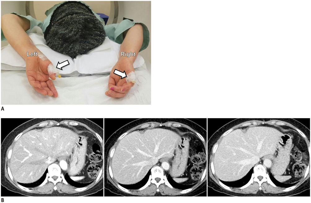
Fig. 1 Placement of 2 small intravenous catheters and dynamic liver CT obtained using them in 61-year-old female. A. Two intravenous catheters (24- and 22-gauge) placed in left thumb and lateral side of right hand (arrows), respectively. B. Arterial- (left), portal- (middle), and delayed- (right) phase contrast-enhanced CT images obtained with 4 mL/s total injection rate. CT = computed tomography
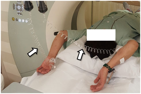
Fig. 2 Simultaneous contrast injection through 2 intravenous catheters. Two coiled intravenous tubing (arrows) are observed, one each connecting one cylinder of dual-head injector to one intravenous catheter (this exemplary patient is not from study cohort).
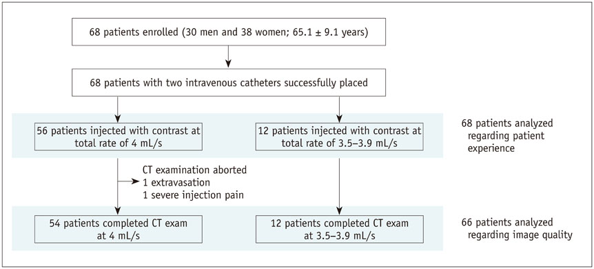
Fig. 3 Patient flow diagram.
Reference
-
1. Fishman EK. CTisus: everything you need to know about computed tomography (CT) & CT scanning. August 30, 2017. Web site. http://www.ctisus.com.2. Springer Healthcare. MDCT.net. August 30, 2017Springer Healthcare. Web site. https://mdct.net.3. Johnson PT, Christensen GM, Fishman EK. I.v. contrast administration with dual source 128-MDCT: a randomized controlled study comparing 18-gauge nonfenestrated and 20-gauge fenestrated catheters for catheter placement success, infusion rate, image quality, and complications. AJR Am J Roentgenol. 2014; 202:1166–1170.
Article4. Schima W, Hammerstingl R, Catalano C, Marti-Bonmati L, Rummeny EJ, Montero FT, et al. Quadruple-phase MDCT of the liver in patients with suspected hepatocellular carcinoma: effect of contrast material flow rate. AJR Am J Roentgenol. 2006; 186:1571–1579.
Article5. Cho JS, Kwag JG, Oh YR, Han SD, Song CJ. Detection and characterization of hepatocellular carcinoma: value of dynamic CT during the arterial dominant phase with uniphasic contrast medium injection. J Comput Assist Tomogr. 1996; 20:128–134.
Article6. Behrendt FF, Bruners P, Keil S, Plumhans C, Mahnken AH, Stanzel S, et al. Impact of different vein catheter sizes for mechanical power injection in CT: in vitro evaluation with use of a circulation phantom. Cardiovasc Intervent Radiol. 2009; 32:25–31.
Article7. Wienbeck S, Fischbach R, Kloska SP, Seidensticker P, Osada N, Heindel W, et al. Prospective study of access site complications of automated contrast injection with peripheral venous access in MDCT. AJR Am J Roentgenol. 2010; 195:825–829.
Article8. Nicola R, Shaqdan KW, Aran S, Prabhakar AM, Singh AK, Abujudeh HH. Contrast media extravasation of computed tomography and magnetic resonance imaging: management guidelines for the radiologist. Curr Probl Diagn Radiol. 2016; 45:161–164.
Article9. Pacheco Compaña FJ, Gago Vidal B, Méndez Díaz C. [Extravasation of contrast media at the puncture site: strategies for managment]. Radiologia. 2014; 56:295–302.10. Sbitany H, Koltz PF, Mays C, Girotto JA, Langstein HN. CT contrast extravasation in the upper extremity: strategies for management. Int J Surg. 2010; 8:384–386.
Article11. Sandler KL, Markham LW, Mah ML, Byrum EP, Williams JR. Optimizing CT angiography in patients with Fontan physiology: single-center experience of dual-site power injection. Clin Radiol. 2014; 69:e562–e567.
Article12. Prabhu SP, Mahmood S, Sena L, Lee EY. MDCT evaluation of pulmonary embolism in children and young adults following a lateral tunnel Fontan procedure: optimizing contrastenhancement techniques. Pediatr Radiol. 2009; 39:938–944.
Article13. Shuman WP, Chan KT, Busey JM, Mitsumori LM, Choi E, Koprowicz KM, et al. Standard and reduced radiation dose liver CT images: adaptive statistical iterative reconstruction versus model-based iterative reconstruction-comparison of findings and image quality. Radiology. 2014; 273:793–800.
Article14. Park SH, Kim PN, Kim KW, Lee SW, Yoon SE, Park SW, et al. Macrovesicular hepatic steatosis in living liver donors: use of CT for quantitative and qualitative assessment. Radiology. 2006; 239:105–112.
Article15. Shuman WP, O'Malley RB, Busey JM, Ramos MM, Koprowicz KM. Prospective comparison of dual-energy CT aortography using 70% reduced iodine dose versus single-energy CT aortography using standard iodine dose in the same patient. Abdom Radiol. 2017; 42:759–765.
Article16. Lv P, Liu J, Chai Y, Yan X, Gao J, Dong J. Automatic spectral imaging protocol selection and iterative reconstruction in abdominal CT with reduced contrast agent dose: initial experience. Eur Radiol. 2017; 27:374–383.
Article17. Ma CL, Chen XX, Lei YX, Zhang XR, Jia YJ, Tian X, et al. Clinical value of dual-energy spectral imaging with adaptive statistical iterative reconstruction for reducing contrast medium dose in CT portal venography: in comparison with standard 120-kVp imaging protocol. Br J Radiol. 2016; 89:20151022.
Article18. Lee JW, Lee G, Lee NK, Moon JI, Ju YH, Suh YJ, et al. Effectiveness of adaptive statistical iterative reconstruction for 64-slice dual-energy computed tomography pulmonary angiography in patients with a reduced iodine load: comparison with standard computed tomography pulmonary angiography. J Comput Assist Tomogr. 2016; 40:777–783.19. Iyama Y, Nakaura T, Yokoyama K, Kidoh M, Harada K, Tokuyasu S, et al. Impact of knowledge-based iterative model reconstruction in abdominal dynamic CT with low tube voltage and low contrast dose. AJR Am J Roentgenol. 2016; 206:687–693.
Article20. Chung YE, You JS, Lee HJ, Lim JS, Lee HS, Baek SE, et al. Possible contrast media reduction with low keV monoenergetic images in the detection of focal liver lesions: a dual-energy CT animal study. PLoS One. 2015; 10:e0133170.
Article21. Noda Y, Kanematsu M, Goshima S, Kondo H, Watanabe H, Kawada H, et al. Reduction of iodine load in CT imaging of pancreas acquired with low tube voltage and an adaptive statistical iterative reconstruction technique. J Comput Assist Tomogr. 2014; 38:714–720.
Article22. Chen CM, Chu SY, Hsu MY, Liao YL, Tsai HY. Low-tube-voltage(80 kVp) CT aortography using 320-row volume CT with adaptive iterative reconstruction: lower contrast medium and radiation dose. Eur Radiol. 2014; 24:460–468.23. Nakaura T, Nakamura S, Maruyama N, Funama Y, Awai K, Harada K, et al. Low contrast agent and radiation dose protocol for hepatic dynamic CT of thin adults at 256-detector row CT: effect of low tube voltage and hybrid iterative reconstruction algorithm on image quality. Radiology. 2012; 264:445–454.
Article24. Goo HW, Goo JM. Dual-energy CT: new horizon in medical imaging. Korean J Radiol. 2017; 18:555–569.
Article
- Full Text Links
-
- Actions
-
Cited
- CITED
-
- Close
- Share
-
- Similar articles
-
- Intravenous contrast media application using cone-beam computed tomography in a rabbit model
- Insertion and Management of Central Venous Catheters
- Roentgenographic Confirmation of Central Venous Catheter Tips
- Placement of Central Venous Access via Subclavian Vein under Fluoroscopic Guidance with Intravenous Contrast Injection
- Massive Intraventricular Air Embolism after Contrast-enhanced CT: Report of Two Cases
