Intravenous contrast media application using cone-beam computed tomography in a rabbit model
- Affiliations
-
- 1Department of Oral and Maxillofacial Radiology, School of Dentistry, Kyung Hee University, Seoul, Korea. hehan@khu.ac.kr
- KMID: 2116789
- DOI: http://doi.org/10.5624/isd.2015.45.1.31
Abstract
- PURPOSE
This study was performed to evaluate the feasibility of visualizing soft tissue lesions and vascular structures using contrast-enhanced cone-beam computed tomography (CE-CBCT) after the intravenous administration of a contrast medium in an animal model.
MATERIALS AND METHODS
CBCT was performed on six rabbits after a contrast medium was administered using an injection dose of 2 mL/kg body weight and an injection rate of 1 mL/s via the ear vein or femoral vein under general anesthesia. Artificial soft tissue lesions were created through the transplantation of autologous fatty tissue into the salivary gland. Volume rendering reconstruction, maximum intensity projection, and multiplanar reconstruction images were reconstructed and evaluated in order to visualize soft tissue contrast and vascular structures.
RESULTS
The contrast enhancement of soft tissue was possible using all contrast medium injection parameters. An adequate contrast medium injection parameter for facilitating effective CE-CBCT was a 5-mL injection before exposure combined with a continuous 5-mL injection during scanning. Artificial soft tissue lesions were successfully created in the animals. The CE-CBCT images demonstrated adequate opacification of the soft tissues and vascular structures.
CONCLUSION
Despite limited soft tissue resolution, the opacification of vascular structures was observed and artificial soft tissue lesions were visualized with sufficient contrast to the surrounding structures. The vascular structures and soft tissue lesions appeared well delineated in the CE-CBCT images, which was probably due to the superior spatial resolution of CE-CBCT compared to other techniques, such as multislice computed tomography.
MeSH Terms
Figure
-

Fig. 1 A catheter is positioned for intravenous injection in New Zealand white rabbit.

Fig. 2 A specially designed equipment is used for immobilization of the rabbit during the procedures.
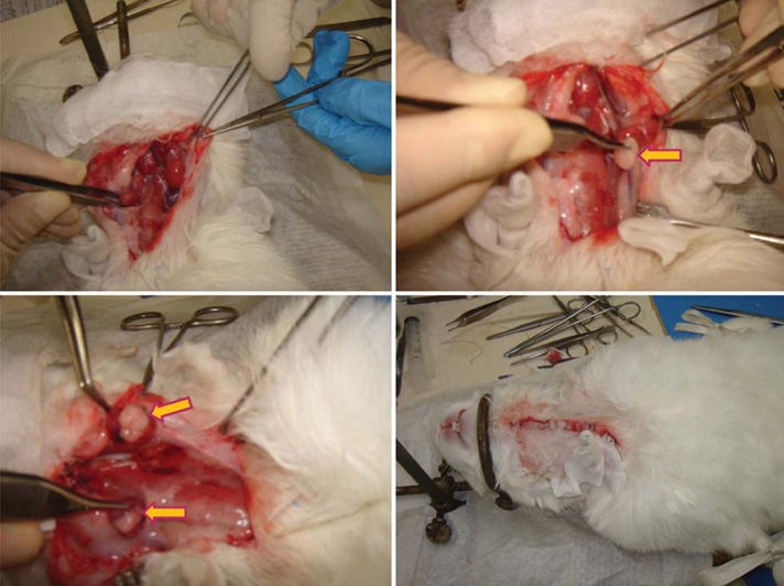
Fig. 3 The photographs show the procedure of an artificial lesion creation in submandibular gland of the rabbit.
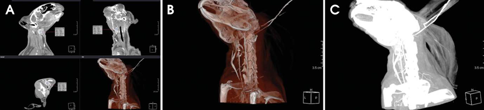
Fig. 4 Using the multiplanar reconstruction (A), volume rendering reconstruction (B), and maximum intensity projection (C) images, the measurements for the submandibular gland are performed.
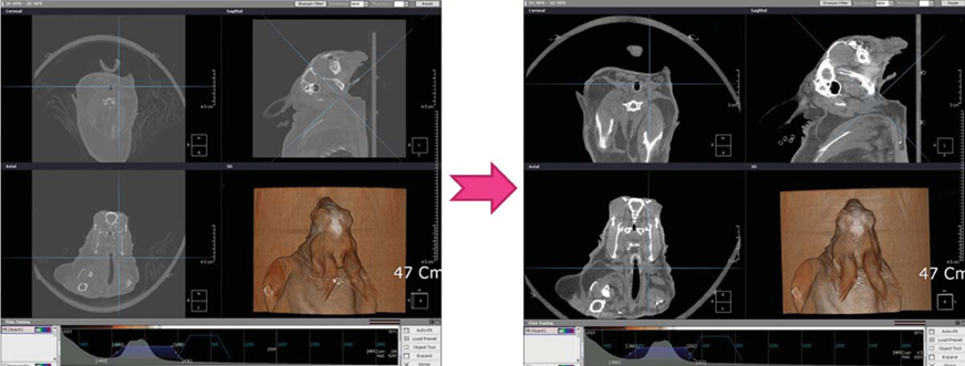
Fig. 5 The window level and width of CBCT images are set at 0 and 1,000, respectively, in order to clarify the submandibular gland and the adjacent fatty space
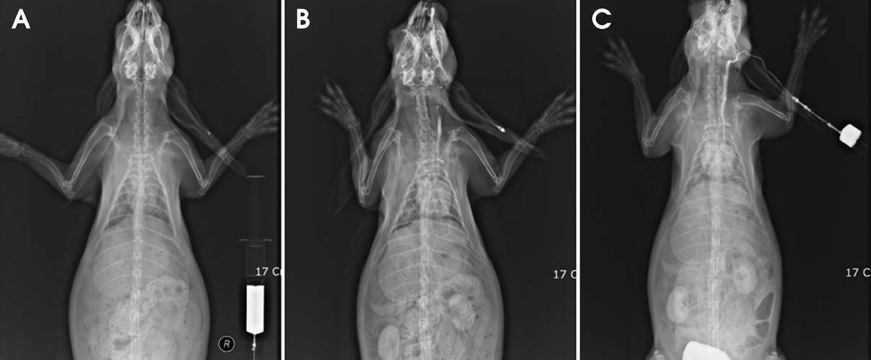
Fig. 6 Effect of time-dependent intravenous contrast enhancement. A. before enhancement. B. 30 seconds after intravenous injection of 10 mL contrast medium. C. combination of continuous injection of 5 mL with 30 seconds delay time after 5 mL injection
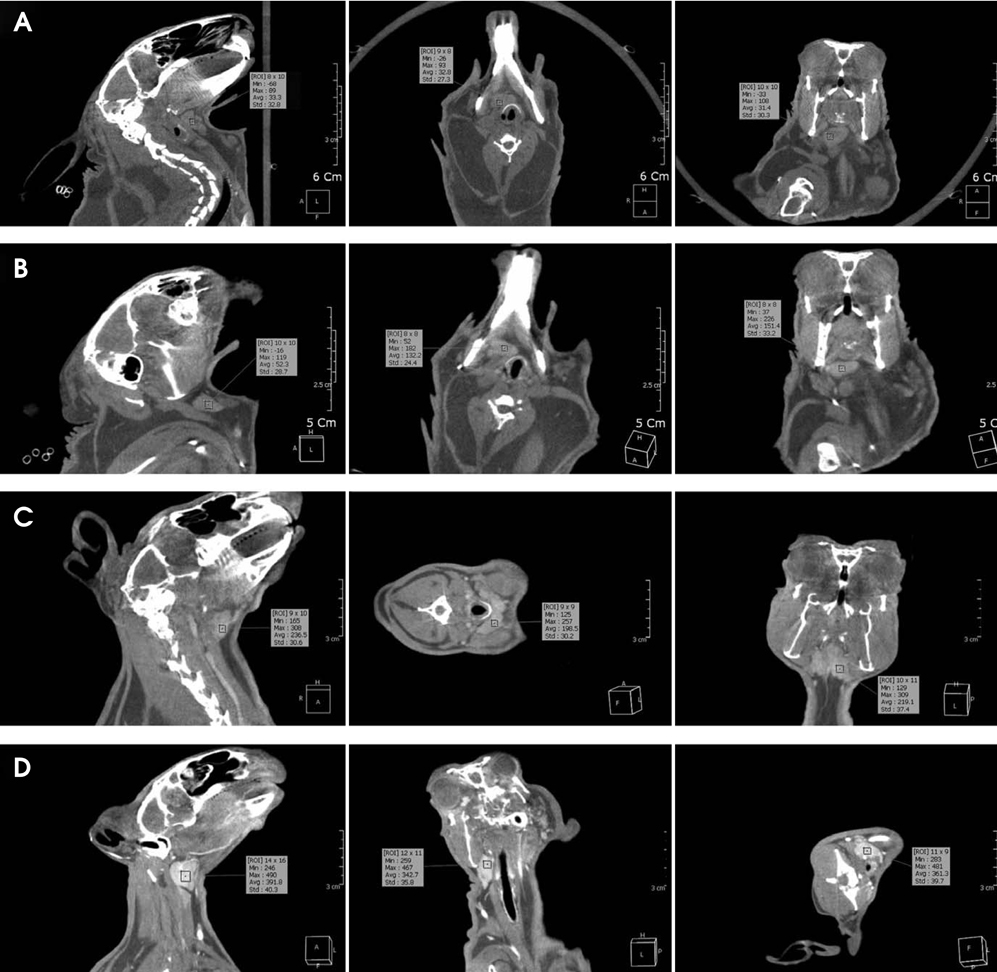
Fig. 7 The contrast density is measured on the non-enhanced CBCT images (A), type 1 enhanced CBCT image (B), type 2 enhanced CBCT image (C), and type 3 enhanced CBCT image (D).

Fig. 8 The graph shows the contrast density in CBCT according to the study protocol of the contrast media administration for contrast enhancement.
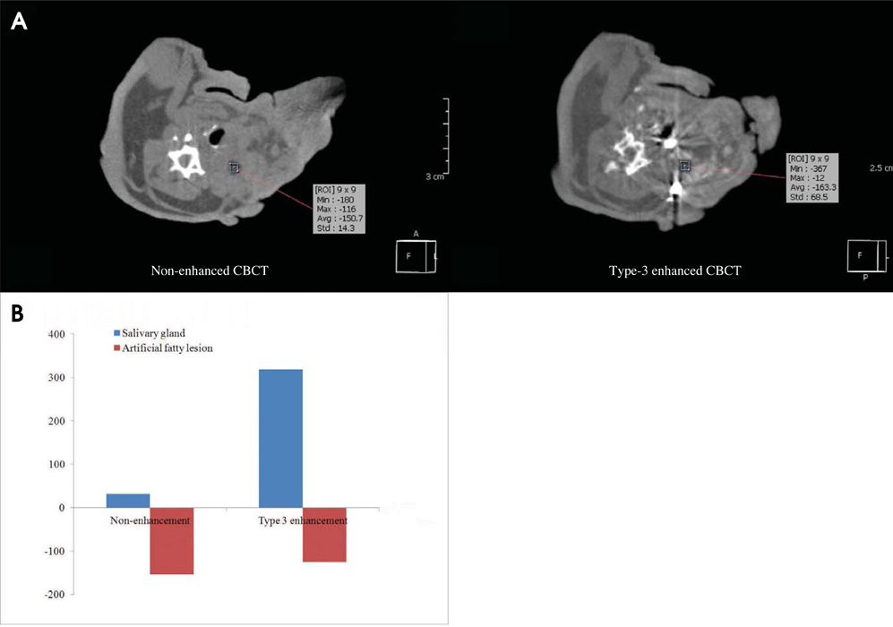
Fig. 9 A. The CBCT images show the difference of value of contrast density before and after contrast media administration for the artificial lesion in salivary gland by transplantation of autogenous fatty tissue. B. The graph shows the comparison of contrast density between non-enhancement and type 3 contrast enhancement in salivary gland and artificial fatty lesion on the CBCT image.
Reference
-
1. Kalender WA, Kyriakou Y. Flat-detector computed tomography (FD-CT). Eur Radiol. 2007; 17:2767–2779.
Article2. Kalender WA. The use of flat-panel detectors for CT imaging. Radiologe. 2003; 43:379–387.3. Scarfe WC, Farman AG, Sukovic P. Clinical applications of cone-beam computed tomography in dental practice. J Can Dent Assoc. 2006; 72:75–80.4. Kapila S, Conley RS, Harrell WE Jr. The current status of cone beam computed tomography imaging in orthodontics. Dentomaxillofac Radiol. 2011; 40:24–34.
Article5. Yu JJ, Kim GT, Choi YS, Hwang EH, Paek J, Kim SH, et al. Accuracy of a cone beam computed tomography-guided surgical stent for orthodontic mini-implant placement. Angle Orthod. 2012; 82:275–283.
Article6. Kim SH, Kang SM, Choi YS, Kook YA, Chung KR, Huang JC. Cone-beam computed tomography evaluation of mini-implants after placement: is root proximity a major risk factor for failure? Am J Orthod Dentofacial Orthop. 2010; 138:264–276.
Article7. Tyndall DA, Rathore S. Cone-beam CT diagnostic applications: caries, periodontal bone assessment, and endodontic applications. Dent Clin North Am. 2008; 52:825–841.
Article8. Kim GT, Kim SH, Choi YS, Park YJ, Chung KR, Suk KE, et al. Cone-beam computed tomography evaluation of orthodontic miniplate anchoring screws in the posterior maxilla. Am J Orthod Dentofacial Orthop. 2009; 136:628.e1–628.e10.
Article9. Choi HS, Kim GT, Choi YS, Hwang EH. Surgical stent for dental implant using cone beam CT images. Korean J Oral Maxillofac Radiol. 2010; 40:171–178.10. Kim SH, Choi YS, Hwang EH, Chung KR, Kook YA, Nelson G. Surgical positioning of orthodontic mini-implants with guides fabricated on models replicated with cone-beam computed tomography. Am J Orthod Dentofacial Orthop. 2007; 131:S82–S89.
Article11. Vercruyssen M, Jacobs R, Van Assche N, van Steenberghe D. The use of CT scan based planning for oral rehabilitation by means of implants and its transfer to the surgical field: a critical review on accuracy. J Oral Rehabil. 2008; 35:454–474.
Article12. Jin JY, Ren L, Liu Q, Kim J, Wen N, Guan H, et al. Combining scatter reduction and correction to improve image quality in cone-beam computed tomography (CBCT). Med Phys. 2010; 37:5634–5644.
Article13. Miracle AC, Mukherji SK. Conebeam CT of the head and neck, part 1: physical principles. AJNR Am J Neuroradiol. 2009; 30:1088–1095.
Article14. Struffert T, Richter G, Engelhorn T, Doelken M, Goelitz P, Kalender WA, et al. Visualisation of intracerebral haemorrhage with flat-detector CT compared to multislice CT: results in 44 cases. Eur Radiol. 2009; 19:619–625.
Article15. Doelken M, Struffert T, Richter G, Engelhorn T, Nimsky C, Ganslandt O, et al. Flat-panel detector volumetric CT for visualization of subarachnoid hemorrhage and ventricles: preliminary results compared to conventional CT. Neuroradiology. 2008; 50:517–523.
Article16. Naitoh M, Nakahara K, Suenaga Y, Gotoh K, Kondo S, Ariji E. Comparison between cone-beam and multislice computed tomography depicting mandibular neurovascular canal structures. Oral Surg Oral Med Oral Pathol Oral Radiol Endod. 2010; 109:e25–e31.
Article17. Watanabe H, Honda E, Tetsumura A, Kurabayashi T. A comparative study for spatial resolution and subjective image characteristics of a multi-slice CT and a cone-beam CT for dental use. Eur J Radiol. 2011; 77:397–402.
Article18. Engelhorn T, Struffert T, Richter G, Doelken M, Ganslandt O, Kalender W, et al. Flat panel detector angiographic CT in the management of aneurysmal rupture during coil embolization. AJNR Am J Neuroradiol. 2008; 29:1581–1584.
Article19. Meyer BC, Frericks BB, Albrecht T, Wolf KJ, Wacker FK. Contrast-enhanced abdominal angiographic CT for intra-abdominal tumor embolization: a new tool for vessel and soft tissue visualization. Cardiovasc Intervent Radiol. 2007; 30:743–749.
Article20. Meyer BC, Frericks BB, Voges M, Borchert M, Martus P, Justiz J, et al. Visualization of hypervascular liver lesions during TACE: comparison of angiographic C-arm CT and MDCT. AJR Am J Roentgenol. 2008; 190:W263–W269.
Article21. Struffert T, Doelken M, Adamek E, Schwarz M, Engelhorn T, Kloska S, et al. Flat-detector computed tomography with intravenous contrast material application in experimental aneurysms: comparison with multislice CT and conventional angiography. Acta Radiol. 2010; 51:431–437.
Article22. Kallmes DF, Helm GA, Hudson SB, Altes TA, Do HM, Mandell JW, et al. Histologic evaluation of platinum coil embolization in an aneurysm model in rabbits. Radiology. 1999; 213:217–222.
Article23. Qu XM, Li G, Ludlow JB, Zhang ZY, Ma XC. Effective radiation dose of ProMax 3D cone-beam computerized tomography scanner with different dental protocols. Oral Surg Oral Med Oral Pathol Oral Radiol Endod. 2010; 110:770–776.
Article24. Carrafiello G, Dizonno M, Colli V, Strocchi S, Pozzi Taubert S, Leonardi A, et al. Comparative study of jaws with multislice computed tomography and cone-beam computed tomography. Radiol Med. 2010; 115:600–611.
Article25. Suomalainen A, Kiljunen T, Käser Y, Peltola J, Kortesniemi M. Dosimetry and image quality of four dental cone beam computed tomography scanners compared with multislice computed tomography scanners. Dentomaxillofac Radiol. 2009; 38:367–378.
Article26. Ritter L, Mischkowski RA, Neugebauer J, Dreiseidler T, Scheer M, Keeve E, et al. The influence of body mass index, age, implants, and dental restorations on image quality of cone beam computed tomography. Oral Surg Oral Med Oral Pathol Oral Radiol Endod. 2009; 108:e108–e116.
Article27. Miyayama S, Yamashiro M, Hattori Y, Orito N, Matsui K, Tsuji K, et al. Efficacy of cone-beam computed tomography during transcatheter arterial chemoembolization for hepatocellular carcinoma. Jpn J Radiol. 2011; 29:371–377.
Article28. Miyayama S, Matsui O, Yamashiro M, Ryu Y, Takata H, Takeda T, et al. Detection of hepatocellular carcinoma by CT during arterial portography using a cone-beam CT technology: comparison with conventional CTAP. Abdom Imaging. 2009; 34:502–506.
Article29. Kyriakou Y, Richter G, Dörfler A, Kalender WA. Neuroradiologic applications with routine C-arm flat panel detector CT: evaluation of patient dose measurements. AJNR Am J Neuroradiol. 2008; 29:1930–1936.
Article30. Struffert T, Hertel V, Kyriakou Y, Krause J, Engelhorn T, Schick B, et al. Imaging of cochlear implant electrode array with flat-detector CT and conventional multislice CT: comparison of image quality and radiation dose. Acta Otolaryngol. 2010; 130:443–452.
Article
- Full Text Links
-
- Actions
-
Cited
- CITED
-
- Close
- Share
-
- Similar articles
-
- Detection of maxillary second molar with two palatal roots using cone beam computed tomography: a case report
- Management of root canal perforation by using cone-beam computed tomography
- Three-dimensional imaging modalities in endodontics
- Transient Orbitofacial Angioedema due to Intravenous Iodinated Contrast Media During Computed Tomography: CT Findings
- Patient radiation dose and protection from cone-beam computed tomography
