Variation of canine vertebral bone architecture in computed tomography
- Affiliations
-
- 1College of Veterinary Medicine, Chonnam National University, Gwangju 61186, Korea. imsono@chonnam.ac.kr
- KMID: 2402705
- DOI: http://doi.org/10.4142/jvs.2018.19.1.145
Abstract
- Focal vertebral bone density changes were assessed in vertebral computed tomography (CT) images obtained from clinically healthy dogs without diseases that affect bone density. The number, location, and density of lesions were determined. A total of 429 vertebral CT images from 20 dogs were reviewed, and 99 focal vertebral changes were identified in 14 dogs. Focal vertebral bone density changes were mainly found in thoracic vertebrae (29.6%) as hyperattenuating (86.9%) lesions. All focal vertebral changes were observed at the vertebral body, except for a single hyperattenuating change in one thoracic transverse process. Among the hyperattenuating changes, multifocal changes (53.5%) were more common than single changes (46.5%). Most of the hypoattenuating changes were single (92.3%). Eight dogs, 40% of the 20 dogs in the study and 61.6% of the 13 dogs showing focal vertebral changes in the thoracic vertebra, had hyperattenuating changes at the 7th or 8th thoracic vertebra. Our results indicate that focal changes in vertebral bone density are commonly identified on vertebral CT images in healthy dogs, and these changes should be taken into consideration on interpretation of CT images.
Keyword
Figure
-
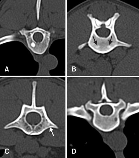
Fig. 1 Transverse computed tomographic images of vertebrae in clinically healthy dogs showing focal attenuation changes; hyperattenuating change as single (A) and multiple (B, arrows) lesions in vertebral bodies, a single hyperattenuating change (C, arrow) at the junction between the left transverse vertebra and the vertebral body, and a single hypoattenuating lesion (D).
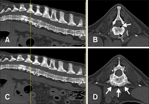
Fig. 2 Transverse computed tomographic (CT) images (B and D) with corresponding reference (dotted lines) on the reconstructed sagittal planes (A and C) of vertebrae in a dog with spondylosis deformans. On transverse images of the 1st lumbar vertebra (B), a solitary hyperattenuating lesion (black arrow) in the vertebral body was observed, and continuation of the hyperattenuating lesion with spondylosis deformans was not observed in adjacent CT slices. A gas (white arrow) accumulation within the vertebral body, a vacuum phenomenon, was incidentally found. Transverse images of the 2nd lumbar vertebra (D) revealed osteoproductive change (white arrows) in the ventral aspect of the vertebral body related with spondylosis deformans.
Reference
-
1. Algra PR, Heimans JJ, Valk J, Nauta JJ, Lachniet M, Van Kooten B. Do metastases in vertebrae begin in the body or the pedicles? Imaging study in 45 patients. AJR Am J Roentgenol. 1992; 158:1275–1279.
Article2. Araki M, Hashimoto K, Kawashima S, Matsumoto K, Akiyama Y. Radiographic features of enostosis determined with limited cone-beam computed tomography in comparison with rotational panoramic radiography. Oral Radiology. 2006; 22:27–33.
Article3. Betbeze C, McLaughlin R. Canine diskospondylitis: its etiology, diagnosis, and treatment. Vet Med Samll Anim Clinician. 2002; 97:673–682.4. Cheon H, Choi W, Lee Y, Lee D, Kim J, Kang JH, Na K, Chang J, Chang D. Assessment of trabecular bone mineral density using quantitative computed tomography in normal cats. J Vet Med Sci. 2012; 74:1461–1467.
Article5. Drost WT, Love NE, Berry CR. Comparison of radiography, myelography and computed tomography for the evaluation of canine vertebral and spinal cord tumors in sixteen dogs. Vet Radiol Ultrasound. 1996; 37:28–33.
Article6. Ertungealp E, Seyisoglu H, Erel CT, Senturk LM, Gezer A. Changes in bone mineral density with age, menopausal status and body mass index in Turkish women. Climacteric. 1999; 2:45–51.
Article7. Evans HE, De Lahunta A. The skeleton. In : Evans HE, De Lahunta A, editors. Miller's Anatomy of the Dog. 4th ed. St. Louis: Elsevier;2013. p. 113–125.8. Goedegebuure SA. Secondary bone tumours in the dog. Vet Pathol. 1979; 16:520–529.
Article9. Gonzalo-Orden JM, Altónaga JR, Orden MA, Gonzalo JM. Magnetic resonance, computed tomographic and radiologic findings in a dog with discospondylitis. Vet Radiol Ultrasound. 2000; 41:142–144.
Article10. Gossios KJ, Kontoyiannis DS, Dascalogiannaki M, Gourtsoyiannis NC. Uncommon locations of hydatid disease: CT appearances. Eur Radiol. 1997; 7:1303–1308.
Article11. Gould CF, Ly JQ, Lattin GE Jr, Beall DP, Sutcliffe JB 3rd. Bone tumor mimics: avoiding misdiagnosis. Curr Probl Diagn Radiol. 2007; 36:124–141.
Article12. Greenspan A, Steiner G, Knutzon R. Bone island (enostosis): clinical significance and radiologic and pathologic correlations. Skeletal Radiol. 1991; 20:85–90.
Article13. Laredo JD, el Quessar A, Bossard P, Vuillemin-Bodaghi V. Vertebral tumors and pseudotumors. Radiol Clin North Am. 2001; 39:137–163.
Article14. Mazess RB, Barden HS, Drinka PJ, Bauwens SF, Orwoll ES, Bell NH. Influence of age and body weight on spine and femur bone mineral density in U.S. white men. J Bone Miner Res. 1990; 5:645–652.
Article15. McCarthy EF, Dorfman HD. Idiopathic segmental sclerosis of vertebral bodies. Skeletal Radiol. 1982; 9:88–91.
Article16. Reeder MM. Comprehensive lists of roentgen differential diagnosis. In : Reeder MM, Felson B, editors. Reeder and Felson's Gamuts in Radiology. 3rd ed. New York: Springer-Verlag;1993. p. 53–68.17. Rusbridge C, Wheeler SJ, Lamb CR, Page RL, Carmichael S, Brearley MJ, Bjornson AP. Vertebral plasma cell tumors in 8 dogs. J Vet Intern Med. 1999; 13:126–133.
Article18. Rybak LD, Rosenthal DI. Radiological imaging for the diagnosis of bone metastases. Q J Nucl Med. 2001; 45:53–64.19. Sande RD. Radiography, myelography, computed tomography, and magnetic resonance imaging of the spine. Vet Clin North Am Small Anim Pract. 1992; 22:811–831.
Article20. Seiler G, Kinns J, Dennison S, Saunders J, Schwarz T. Vertebral column and spinal cord. In : Schwarz T, Saunders J, editors. Veterinary Computed Tomography. West Sussex: Wiley-Blackwell;2011. p. 209–228.21. Steingruber IE, Bach CM, Wimmer C, Nogler M, Buchberger W. Multisegmental pneumatocysts of the lumbar spine mimic osteolytic lesions. Eur Radiol. 2001; 11:845–848.
Article22. Torricelli P, Martinelli C, Biagini R, Ruggieri P, De Cristofaro R. Radiographic and computed tomographic findings in hydatid disease of bone. Skeletal Radiol. 1990; 19:435–439.
Article23. Valentine BA, McGavin MN. Bone and joints. In : McGavin MD, Zachary JF, editors. Pathological Basis of Veterinary Disease. 4th ed. St. Louis: Elsevier;2007. p. 1044–1052.24. Zotti A, Gianesella M, Gasparinetti N, Zanetti E, Cozzi B. A preliminary investigation of the relationship between the “moment of resistance” of the canine spine, and the frequency of traumatic vertebral lesions at different spinal levels. Res Vet Sci. 2011; 90:179–184.
Article
- Full Text Links
-
- Actions
-
Cited
- CITED
-
- Close
- Share
-
- Similar articles
-
- Evaluation of mandibular cortical bone thickness for placement of temporary anchorage devices (TADs)
- Monostotic Paget's Disease of the Lumbar Spine Mimicking Vertebral Metastasis: A Case Report
- Novel vertebral computed tomography indices in normal and spinal disorder dogs
- Variation in the vertebral levels of the origins of the abdominal aorta branches: a retrospective imaging study
- Relationship between trabecular strength and three-dimensional architecture in the pig mandible using microcomputed tomography
