J Vet Sci.
2018 Mar;19(2):296-300. 10.4142/jvs.2018.19.2.296.
Novel vertebral computed tomography indices in normal and spinal disorder dogs
- Affiliations
-
- 1Institute of Animal Medicine, College of Veterinary Medicine, Gyeongsang National University, Jinju 30488, Korea. lhc@gnu.ac.kr
- KMID: 2407629
- DOI: http://doi.org/10.4142/jvs.2018.19.2.296
Abstract
- This study was carried out to derive and evaluate reference computed tomography (CT)-based indices for normal canine spine. CT and magnetic resonance images were acquired from 12 clinically normal Beagle dogs (normal group) and 50 dogs with 56 spinal disorders (patient group). Image acquisition regions were cervical spine (C2-T1), thoracic spine (T1-T13), and lumbar spine (L1-L7). Measured indices were: the ratios of width to height of the spinal cord including the dura matter (CR) and of the vertebral foramen (FR), and the ratio of the cross-sectional area of the spinal cord to that of the vertebral foramen (CFAR). Reliability analysis was performed to evaluate intermodality agreement. Student's t-tests and receiver operating characteristic curves were used to discriminate the normal and patient groups on CT. Intermodality agreements of the normal and patient groups were acceptable to excellent. The highest discriminating levels of CR at the vertebral body level and the intervertebral disc space level were 1.25 or more and 1.44 or more, respectively. FR and CFAR had the highest discriminating level at the cervical region. This report presents quantitative information on canine spinal morphometry; the obtained indices may be helpful for CT screening of dogs with spinal disorders.
MeSH Terms
Figure
-
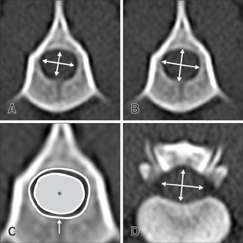
Fig. 1 Method of measuring the variables. The ratios of width to height of the spinal cord (A and D) and of the vertebral foramen (B) were obtained. (C) The ratio of cross-sectional area of the spinal cord (asterisk) to that of the vertebral foramen (arrow) was measured. All measurements were obtained at the vertebral body level (A–C) or intervertebral disc space level (D).
Reference
-
1. Arndt WF 3rd, Truax AL, Barnett FM, Simmons GE, Brown DC. MR diagnosis of bone contusions of the knee: comparison of coronal T2-weighted fast spin-echo with fat saturation and fast spin-echo STIR images with conventional STIR images. AJR Am J Roentgenol. 1996; 166:119–124.
Article2. Benigni L, Lamb CR. Comparison of fluid-attenuated inversion recovery and T2-weighted magnetic resonance images in dogs and cats with suspected brain disease. Vet Radiol Ultrasound. 2005; 46:287–292.
Article3. Drost W, Love NE, Berry CR. Comparison of radiography, myelography and computed tomography for the evaluation of canine vertebral and spinal cord tumors in sixteen dogs. Vet Radiol Ultrasound. 1996; 37:28–33.
Article4. Dymarkowski S, Ni Y, Miao Y, Bogaert J, Rademakers F, Bosmans H, Marchal G. Value of T2-weighted magnetic resonance imaging early after myocardial infarction in dogs: comparison with bis-gadolinium-mesoporphyrin enhanced T1-weighted magnetic resonance imaging and functional data from cine magnetic resonance imaging. Invest Radiol. 2002; 37:77–85.
Article5. Feeney DA, Evers P, Fletcher TF, Hardy RM, Wallace LJ. Computed tomography of the normal canine lumbosacral spine: a morphologic perspective. Vet Radiol Ultrasound. 1996; 37:399–411.
Article6. Ferreira VM, Piechnik SK, Dall'Armellina E, Karamitsos TD, Francis JM, Choudhury RP, Friedrich MG, Robson MD, Neubauer S. Non-contrast T1-mapping detects acute myocardial edema with high diagnostic accuracy: a comparison to T2-weighted cardiovascular magnetic resonance. J Cardiovasc Magn Reson. 2012; 14:42.
Article7. Fourie SL, Kirberger RM. Relationship of cervical spinal cord diameter to vertebral dimensions: a radiographic study of normal dogs. Vet Radiol Ultrasound. 1999; 40:137–143.
Article8. Hara Y, Tagawa M, Ejima H, Orima H, Fujita M. Usefulness of computed tomography after myelography for surgery on dogs with cervical intervertebral disc protrusion. J Vet Med Sci. 1994; 56:791–794.
Article9. House PM, Lanz M, Holst B, Martens T, Stodieck S, Huppertz HJ. Comparison of morphometric analysis based on T1- and T2-weighted MRI data for visualization of focal cortical dysplasia. Epilepsy Res. 2013; 106:403–409.
Article10. Kalita J, Misra UK. Comparison of CT scan and MRI findings in the diagnosis of Japanese encephalitis. J Neurol Sci. 2000; 174:3–8.
Article11. Kidwell CS, Chalela JA, Saver JL, Starkman S, Hill MD, Demchuk AM, Butman JA, Patronas N, Alger JR, Latour LL, Luby ML, Baird AE, Leary MC, Tremwel M, Ovbiagele B, Fredieu A, Suzuki S, Villablanca JP, Davis S, Dunn B, Todd JW, Ezzeddine MA, Haymore J, Lynch JK, Davis L, Warach S. Comparison of MRI and CT for detection of acute intracerebral hemorrhage. JAMA. 2004; 292:1823–1830.
Article12. Kim B, Semelka RC, Ascher SM, Chalpin DB, Carroll PR, Hricak H. Bladder tumor staging: comparison of contrast-enhanced CT, T1- and T2-weighted MR imaging, dynamic gadolinium-enhanced imaging, and late gadolinium-enhanced imaging. Radiology. 1994; 193:239–245.
Article13. Mirowitz SA, Apicella P, Reinus WR, Hammerman AM. MR imaging of bone marrow lesions: relative conspicuousness on T1-weighted, fat-suppressed T2-weighted, and STIR images. AJR Am J Roentgenol. 1994; 162:215–221.
Article14. Morgan JP, Atilola M, Bailey CS. Vertebral canal and spinal cord mensuration: a comparative study of its effect on lumbosacral myelography in the dachshund and German shepherd dog. J Am Vet Med Assoc. 1987; 191:951–957.15. Ross MR, Schomer DF, Chappell P, Enzmann DR. MR imaging of head and neck tumors: comparison of T1-weighted contrast-enhanced fat-suppressed images with conventional T2-weighted and fast spin-echo T2-weighted images. AJR Am J Roentgenol. 1994; 163:173–178.
Article16. Schellhas KP, Wilkes CH. Temporomandibular joint inflammation: comparison of MR fast scanning with T1- and T2-weighted imaging techniques. AJR Am J Roentgenol. 1989; 153:93–98.
Article17. Seo E, Choi J, Choi M, Yoon J. Computed tomographic evaluation of cervical vertebral canal and spinal cord morphometry in normal dogs. J Vet Sci. 2014; 15:187–193.
Article18. Stanley JH, Schabel SI, Frey GD, Hungerford GD. Quantitative analysis of the cervical spinal canal by computed tomography. Neuroradiology. 1986; 28:139–143.
Article
- Full Text Links
-
- Actions
-
Cited
- CITED
-
- Close
- Share
-
- Similar articles
-
- Computed tomographic evaluation of cervical vertebral canal and spinal cord morphometry in normal dogs
- Variation of canine vertebral bone architecture in computed tomography
- CT myelography of the thoraco-lumbar spine in 8 dogs with degenerative myelopathy
- Evaluation of gallbladder and common bile duct size and appearance by computed tomography in dogs
- Is sheep lumbar spine a suitable alternative model for human spinal researches? Morphometrical comparison study
