Radiographic and computed tomography monitoring of a fractured needle fragment in the mandibular branch
- Affiliations
-
- 1Department of Dentistry, Pontificial Catholic University of Minas Gerais, Belo Horizonte, Brazil. contato@misabel.com.br
- KMID: 2391409
- DOI: http://doi.org/10.5624/isd.2017.47.1.63
Abstract
- Some complications can arise with the usage of local anesthesia for dental procedures, including the fracture of needles in the patient. This is a rare incident, usually caused by the patient's sudden movements during anesthetic block. Its complications are not common, but can include pain, trismus, inflammation in the region, difficulty in swallowing, and migration of the object, which is the least common but has the ability to cause more serious damage to the patient. This report describes a case in which, after the fracture of the anesthetic needle used during alveolar nerve block for exodontia of the left mandibular third molar, the fragment moved significantly in the first 2 months, before stabilizing after the third month of radiographic monitoring.
Keyword
MeSH Terms
Figure
-
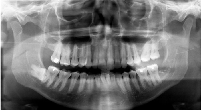
Fig. 1 Initial panoramic radiographs. The fractured needle is indicated with arrows in the area of the ascending branch of the mandible.
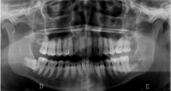
Fig. 2 Panoramic radiograph after 15 days. Notice the fractured needle moving away from the area of the ascending branch of the mandible.
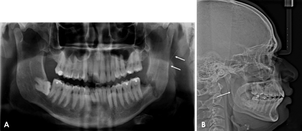
Fig. 3 Panoramic radiograph (A) and lateral cephalography (B) show the foreign object (white arrows) at 1 month of monitoring.

Fig. 4 An axial cone-beam computed tomographic image (bone window) shows the fractured needle in the region of the medial pterygoid muscle.
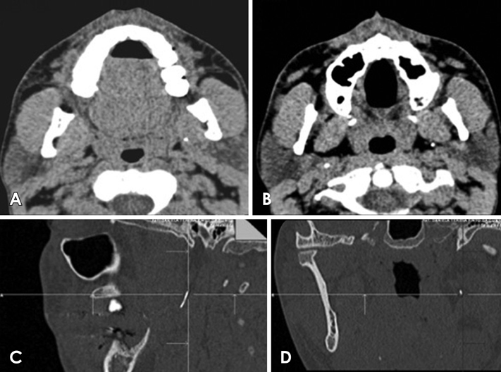
Fig. 5 Multislice computed tomography with 64 detectors. (A and B) The axial images (soft-tissue window) show the foreign body; the top part of the needle was located in the parapharyngeal space and the bottom part in the medial pterygoid muscle. Notice the loss of anatomical fat structures with the formation of granulomatous tissue. (C and D) Sagittal and coronal planes, respectively, which were useful for localizing the needle at 1 A B month of monitoring.
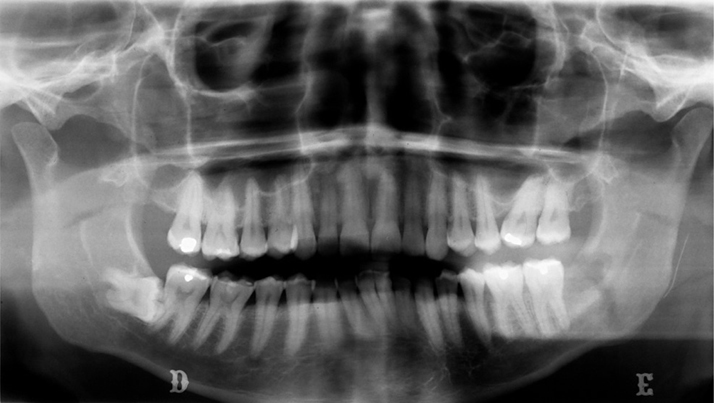
Fig. 6 Panoramic radiography shows the fractured needle, which was located in the posterior area of the ascending branch of the mandible at 2 months of monitoring.
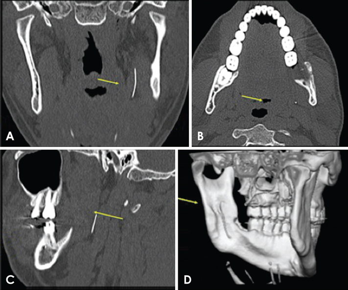
Fig. 7 Multislice computed tomography with 64 detectors. The coronal (A), axial (B), and sagittal planes (C) and 3-dimensional reconstructed (D) images show the foreign body (yellow arrows); the top part of the needle was located in the medial pterygoid muscle and the bottom part in the parapharyngeal space, at 2 months of monitoring.
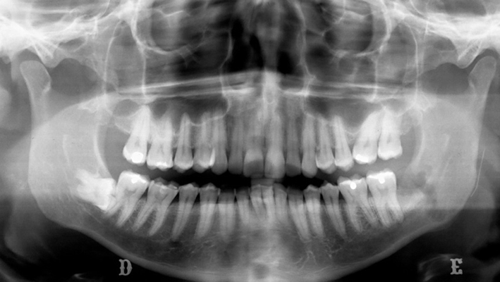
Fig. 8 Panoramic radiograph showing the stabilized fragment at 3 months of monitoring.
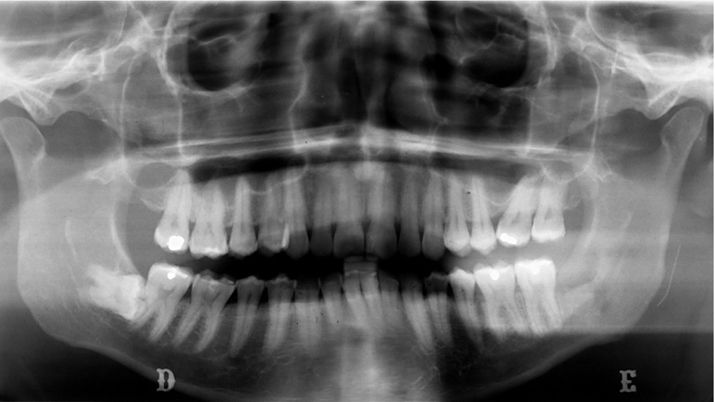
Fig. 9 Panoramic radiograph taken 2 years after the fracture, showing its stable position.
Reference
-
1. Augello M, von Jackowski J, Grätz KW, Jacobsen C. Needle breakage during local anesthesia in the oral cavity - a retrospective of the last 50 years with guidelines for treatment and prevention. Clin Oral Investig. 2011; 15:3–8.2. Bedrock RD, Skigen A, Dolwick MF. Retrieval of a broken needle in the pterygomandibular space. J Am Dent Assoc. 1999; 130:685–687.
Article3. Chrcanovic BR, Menezes Junior DC, Custodio AL. Complication of local dental anesthesia - a broken needle in the pterygomandibular space. Braz J Oral Sci. 2009; 8:159–162.4. Pogrel MA. Broken local anesthetic needles: a case series of 16 patients, with recommendations. J Am Dent Assoc. 2009; 140:1517–1522.5. Ethunandan M, Tran AL, Anand R, Bowden J, Seal MT, Brennan PA. Needle breakage following inferior alveolar nerve block: implications and management. Br Dent J. 2007; 202:395–397.
Article6. Faura-Solé M, Sánchez-Garcés MA, Berini-Aytes L, Gay-Escoda C. Broken anesthetic injection needles: report of 5 cases. Quintessence Int. 1999; 30:461–465.7. Zeltser R, Cohen C, Casap N. The implications of a broken needle in the pterygomandibular space: clinical guidelines for prevention and retrieval. Pediatr Dent. 2002; 24:153–156.8. Kim JH, Moon SY. Removal of a broken needle using three-dimensional computed tomography: a case report. J Korean Assoc Oral Maxillofac Surg. 2013; 39:251–253.
Article9. Nezafati S, Shahi S. Removal of broken dental needle using mobile digital C-arm. J Oral Sci. 2008; 50:351–353.
Article10. Thompson M, Wright S, Cheng LH, Starr D. Locating broken dental needles. Int J Oral Maxillofac Surg. 2003; 32:642–644.
- Full Text Links
-
- Actions
-
Cited
- CITED
-
- Close
- Share
-
- Similar articles
-
- Fractured needle as an unusual complication of the lingual nerve block: a case report
- Reliability of panoramic radiography in predicting proximity of third molars to the mandibular canal: A comparison using cone-beam computed tomography
- Removal of a broken needle using three-dimensional computed tomography: a case report
- Recurrent simple bone cyst of the mandibular condyle: a case report
- Cone beam computed tomography findings of ectopic mandibular third molar in the mandibular condyle: report of a case
