Imaging Sci Dent.
2011 Sep;41(3):135-137. 10.5624/isd.2011.41.3.135.
Cone beam computed tomography findings of ectopic mandibular third molar in the mandibular condyle: report of a case
- Affiliations
-
- 1Department of Oral and Maxillofacial Radiology, School of Dentistry, Chosun University, Gwangju, Korea. hidds@chosun.ac.kr
- KMID: 1449959
- DOI: http://doi.org/10.5624/isd.2011.41.3.135
Abstract
- Impaction of third molar is a common developmental abnormality. However, ectopic impaction of the mandibular third molar in condylar region is an extremely rare condition. This report describes a case of impacted tooth in the mandibular condyle without any associated pathologic condition. Also, this report presents the spatial relationship of the impacted mandibular third molar to the surrounding anatomic structures using cone beam computed tomography.
MeSH Terms
Figure
-
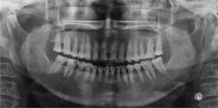
Fig. 1 Panoramic radiograph shows the impacted tooth in the right mandibular condylar head.
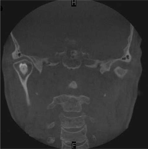
Fig. 2 Coronal CBCT image shows the upward crown position of the impacted tooth. There is no cystic change around impacted tooth.
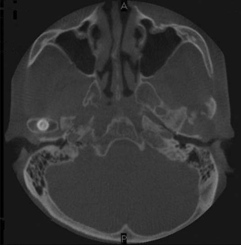
Fig. 3 Axial CBCT image shows the impacted tooth with the proximity to the front cortical bone and middle portion of the mandibular condyle.
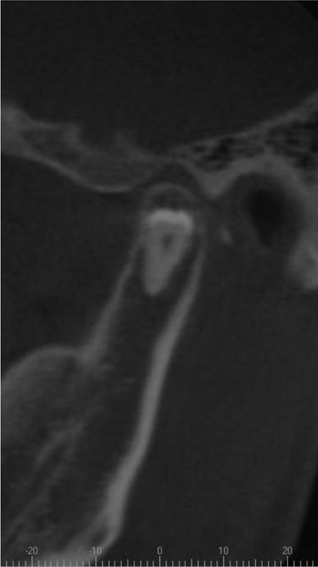
Fig. 4 Sagittal CBCT image shows the fully developed impacted tooth with the proximity to the outer cortical bone of the mandible.
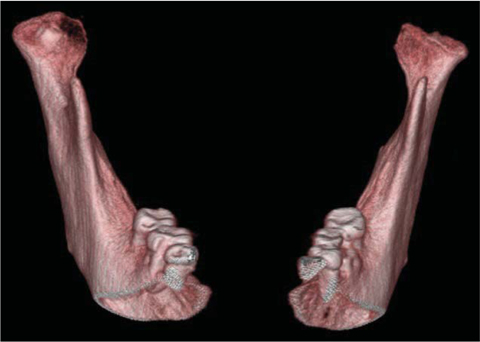
Fig. 5 Frontal view of 3-dimensional volumetric CBCT image shows the crown of the impacted tooth partially covered with bone.
Reference
-
1. Alling CC 3rd, Catone GA. Management of impacted teeth. J Oral Maxillofac Surg. 1993. 51:1 Suppl 1. 3–6.
Article2. Salmerón JI, del Amo A, Plasencia J, Pujol R, Vila CN. Ectopic third molar in condylar region. Int J Oral Maxillofac Surg. 2008. 37:398–400.
Article3. Wang CC, Kok SH, Hou LT, Yang PJ, Lee JJ, Cheng SJ, et al. Ectopic mandibular third molar in the ramus region: report of a case and literature review. Oral Surg Oral Med Oral Pathol Oral Radiol Endod. 2008. 105:155–161.
Article4. Medici A, Raho MT, Anghinoni M. Ectopic third molar in the condylar process: case report. Acta Biomed Ateneo Parmense. 2001. 72:115–118.5. Gadre KS, Waknis P. Intra-oral removal of ectopic third molar in the mandibular condyle. Int J Oral Maxillofac Surg. 2010. 39:294–296.
Article6. Wassouf A, Eyrich G, Lebeda R, Grätz KW. Surgical removal of a dislocated lower third molar from the condyle region: case report. Schweiz Monatsschr Zahnmed. 2003. 113:416–420.7. Park W, Lee JH, Park H, Jung HG, Kim KD. Impacted supernumerary tooth in coronoid process: a case report. Korean J Oral Maxillofac Radiol. 2010. 40:89–91.
- Full Text Links
-
- Actions
-
Cited
- CITED
-
- Close
- Share
-
- Similar articles
-
- Mandibular condyle position in cone beam computed tomography
- Assessment of the relationship between the mandibular third molar and the mandibular canal using panoramic radiograph and cone beam computed tomography
- Recurrent simple bone cyst of the mandibular condyle: a case report
- Positional relationship between mandibular third molar and mandibular canal in cone beam computed tomographs
- Analysis and evaluation of relative positions of mandibular third molar and mandibular canal impacts
