Recurrent simple bone cyst of the mandibular condyle: a case report
- Affiliations
-
- 1Department of Oral and Maxillofacial Radiology, School of Dentistry, and Institute of Oral Bio Science, Chonbuk National University, Jeonju, Korea. beam@jbnu.ac.kr
- KMID: 2167447
- DOI: http://doi.org/10.5624/isd.2013.43.1.49
Abstract
- Cysts of the mandibular condyle are rare and can be difficult to diagnose and treat. Clinically, a simple bone cyst is asymptomatic and often discovered incidentally on routine radiographic examination. This report shows an atypical simple bone cyst occurring in the mandibular condyle showing recurrence after surgical curettage. Radiologically, this lesion involving the mandibular condyle should be distinguished from other similar lesions such as a chondroma, a central giant cell granuloma, and an aneurysmal bone cyst. Radiographic assessment was useful for forecasting the prognosis of a simple bone cyst. Possible reasons for the recurrence were discussed radiographically.
MeSH Terms
Figure
-
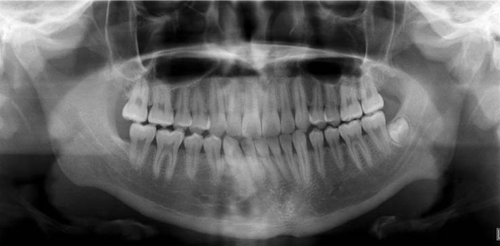
Fig. 1 Panoramic radiograph shows well-defined multilocular radiolucency in the left mandibular condyle.
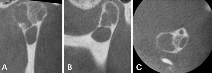
Fig. 2 Coronal (A), sagittal (B), and axial (C) CBCT images show an amorphous multilocular lytic lesion and a scalloped cortical border in some parts.
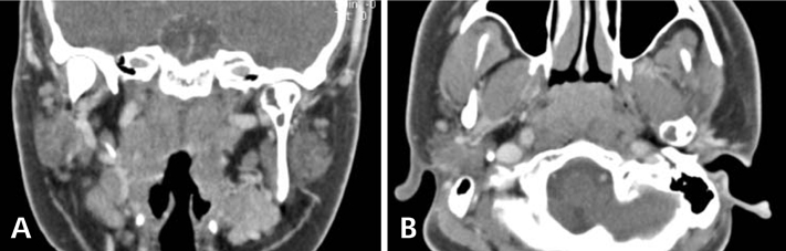
Fig. 3 Coronal (A) and axial (B) enhanced CT scans show a nonenhanced multilocular lesion in the left mandibular condyle.
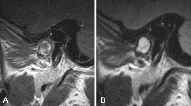
Fig. 4 MRI shows homogeneous intermediate SI on the T1-weighted image (A) and homogeneous high SI on the T2-weighted image (B).
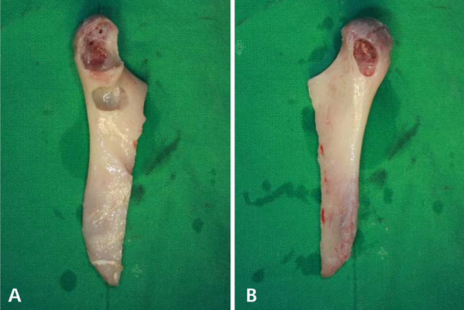
Fig. 5 The eviscerated and curetted mandibular condyle. A. Medial pole. B. Lateral pole.
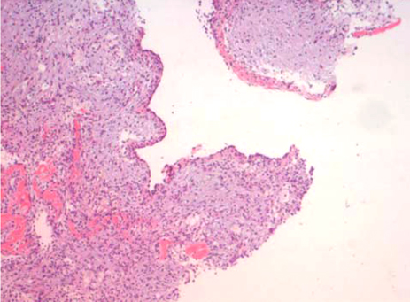
Fig. 6 Microscopic examination shows a cyst without an epithelial lining, which is surrounded by dense fibrous connective tissue and reactive bone (H&E stain, ×100).
Reference
-
1. Forssell K, Forssell H, Happonen RP, Neva M. Simple bone cyst. Review of the literature and analysis of 23 cases. Int J Oral Maxillofac Surg. 1988. 17:21–24.2. Hosseini M. Two atypical solitary bone cysts. Br J Oral Surg. 1979. 16:262–269.
Article3. Cortell-Ballester I, Figueiredo R, Berini-Aytés L, Gay-Escoda C. Traumatic bone cyst: a retrospective study of 21 cases. Med Oral Patol Oral Cir Bucal. 2009. 14:E239–E243.4. Khosla VM. Hemorrhagic bone cyst of mandible. Report of a case. Oral Surg Oral Med Oral Pathol. 1970. 30:723–729.5. Peters RA, Wussow GC. Extravasation cyst of the maxilla. Report of a case. Oral Surg Oral Med Oral Pathol. 1968. 26:742–745.6. Velez I, Siegel MA, Mintz SM, Rolle R. The relationship between idiopathic bone cavity and orthodontic tooth movement: analysis of 44 cases. Dentomaxillofac Radiol. 2010. 39:162–166.
Article7. Robinson M, Canter S, Shuken R. Multiple progressive bone cysts of the mandible and maxilla. Oral Surg Oral Med Oral Pathol. 1967. 23:483–486.
Article8. Persson G. An atypical solitary bone cyst. J Oral Maxillofac Surg. 1985. 43:905–907.
Article9. Gilman RH, Dingman RO. A solitary bone cyst of the mandibular condyle. Plast Reconstr Surg. 1982. 70:610–614.
Article10. Shigematsu H, Fujita K, Watanabe K. Atypical simple bone cyst of the mandible. A case report. Int J Oral Maxillofac Surg. 1994. 23:298–299.
Article11. Tanaka H, Westesson P, Emmings FG, Marashi AH. Simple bone cyst of the mandibular condyle: report of a case. J Oral Maxillofac Surg. 1996. 54:1454–1458.
Article12. Kuttenberger JJ, Farmand M, Stöss H. Recurrence of a solitary bone cyst of the mandibular condyle in a bone graft. A case report. Oral Surg Oral Med Oral Pathol. 1992. 74:550–556.13. Rubin MM, Murphy FJ. Simple bone cyst of the mandibular condyle. J Oral Maxillofac Surg. 1989. 47:1096–1098.
Article14. Magliocca KR, Edwards SP, Helman JI. Traumatic bone cyst of the condylar region: report of 2 cases. J Oral Maxillofac Surg. 2007. 65:1247–1250.
Article15. Jadu FM, Pharoah MJ, Lee L, Baker GI, Allidina A. Central giant cell granuloma of the mandibular condyle: a case report and review of the literature. Dentomaxillofac Radiol. 2011. 40:60–64.
Article16. Yu JJ, Park JH, Kang JH, Kim GT, Choi YS, Hwang EH. Aneurysmal bone cyst of the mandibular condyle with condylar neck fracture. Korean J Oral Maxillofac Radiol. 2009. 39:205–208.17. Motamedi MH. Destructive aneurismal bone cyst of the mandibular condyle: report of a case and review of the literature. J Oral Maxillofac Surg. 2002. 60:1357–1361.18. Damante JH, Da S Guerra EN, Ferreira O Jr. Spontaneous resolution of simple bone cysts. Dentomaxillofac Radiol. 2002. 31:182–186.
Article19. Sapp JP, Stark ML. Self-healing traumatic bone cysts. Oral Surg Oral Med Oral Pathol. 1990. 69:597–602.
Article20. Chapman PJ, Romaniuk K. Traumatic bone cyst of the mandible: regression following aspiration. Int J Oral Surg. 1985. 14:290–294.
Article21. Suei Y, Taguchi A, Nagasaki T, Tanimoto K. Radiographic findings and prognosis of simple bone cysts of the jaws. Dentomaxillofac Radiol. 2010. 39:65–71.
Article22. Feinberg SE, Finkelstein MW, Page HL, Dembo JB. Recurrent "traumatic" bone cysts of the mandible. Oral Surg Oral Med Oral Pathol. 1984. 57:418–422.
Article23. Mathew R, Omami G, Gianoli D, Lurie A. Unusual cone-beam computerized tomography presentation of traumatic (simple) bone cyst: case report and radiographic analysis. Oral Surg Oral Med Oral Pathol Oral Radiol. 2012. 113:410–413.
Article24. Telfer MR, Jones GM, Pell GM, Eveson JW. Primary bone cyst of the mandibular condyle. Br J Oral Maxillofac Surg. 1990. 28:340–343.
Article25. Ogasawara T, Kitagawa Y, Ogawa T, Yamada T, Yamamoto S, Hayashi K. Simple bone cyst of the mandibular condyle with severe osteoarthritis: report of a case. J Oral Pathol Med. 1999. 28:377–380.
Article26. Choi SC, Lee SS, Lee GI. Radiologic study of the traumatic bone cysts. J Korean Acad Oral Maxillofac Radiol. 1994. 24:7–21.27. Oh KR, Park WK, Ko JK, Kim YJ. A study of the traumatic bone cyst. J Korean Acad Oral Maxillofac Radiol. 1997. 27:145–160.28. Suei Y, Tanimoto K, Wada T. Simple bone cyst. Evaluation of contents with conventional radiography and computed tomography. Oral Surg Oral Med Oral Pathol. 1994. 77:296–301.29. Matsuzaki H, Asaumi J, Yanagi Y, Konouchi H, Honda Y, Hisatomi M, et al. MR imaging in the assessment of a solitary bone cyst. Eur J Radiol Extra. 2003. 45:37–42.
Article
- Full Text Links
-
- Actions
-
Cited
- CITED
-
- Close
- Share
-
- Similar articles
-
- Simple bone cyst recurred in adjacent areas: A case report
- Bony window approach for a traumatic bone cyst on the mandibular condyle: a case report with long-term follow-up
- Aneurysmal bone cyst of the mandibular condyle with condylar neck fracture
- Anterior stafne bone cyst mimicking periapical cyst: a case report
- Conservative Treatment of Chronic Suppurative Osteomyelitis on Mandibular Body to Condyle Area: A Case Report
