J Gastric Cancer.
2017 Jun;17(2):180-185. 10.5230/jgc.2017.17.e11.
Gastric Adenocarcinoma Secondary to Primary Gastric Diffuse Large B-cell Lymphoma
- Affiliations
-
- 1Department of Hematology-Oncology, Centre Hospitalier Universitaire Notre Dame des Secours, Byblos, Lebanon. marcel.massoud@gmail.com
- 2Department of Hematology-Oncology, Faculty of Medical Sciences, Holy Spirit University of Kaslik, Jounieh, Lebanon.
- 3Department of Pathology, Centre Hospitalier Universitaire Notre Dame des Secours, Byblos, Lebanon.
- 4Department of Pathology, Faculty of Medical Sciences, Holy Spirit University of Kaslik, Jounieh, Lebanon.
- 5Department of Hematology-Oncology, Hotel Dieu de France Hospital, Universite Saint-Joseph de Beyrouth, Beirut, Lebanon.
- KMID: 2383741
- DOI: http://doi.org/10.5230/jgc.2017.17.e11
Abstract
- Despite the decreasing incidence and mortality from gastric cancer, it remains a major health problem worldwide. Ninety percent of cases are adenocarcinomas. Here, we report a case of gastric adenocarcinoma developed after successful treatment of prior primary gastric diffuse large B-cell lymphoma (DLBCL). Our patient was an elderly man with primary gastric DLBCL in whom complete remission was achieved after R-CHOP (cyclophosphamide, adriamycin, vincristine, prednisolone plus rituximab) chemotherapy. Helicobacter pylori infection persisted despite adequate treatment leading to sustained chronic gastritis. The mean time to diagnose metachronous gastric carcinoma was seven years. We believe that a combination of many risk factors, of which chronic H. pylori infection the most important, led to the development of gastric carcinoma following primary gastric lymphoma. In summary, patients who have been successfully treated for primary gastric lymphoma should be followed up at regular short intervals. H. pylori infection should be diagnosed promptly and treated aggressively.
MeSH Terms
Figure
-

Fig. 1 Abdominal computed tomography scan: 3-cm circumferential wall thickening of the entire stomach, mainly at the fundus and antrum, can be seen. Absence of contrast enhancement.

Fig. 2 Diffuse interstitial infiltration by large lymphoid cells, CD20+, CD10−, and cyclin D1 (H&E, ×100).

Fig. 3 Tumor cells expressing CD20 (immunohistochemistry, ×100).
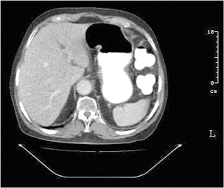
Fig. 4 Computed tomography scan of the abdomen 4 months after completion of chemotherapy showing complete resolution of the previously noted gastric tumor.
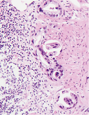
Fig. 5 Glandular proliferation lined with cylindrical epithelial cells with atypical and hyperchromatic nuclei that are compatible with microfoci of invasive gastric adenocarcinoma of the intestinal type (H&E, ×400).
Reference
-
1. Coleman CN, Williams CJ, Flint A, Glatstein EJ, Rosenberg SA, Kaplan HS. Hematologic neoplasia in patients treated for Hodgkin's disease. N Engl J Med. 1977; 297:1249–1252.2. Ioannidis O, Sekouli A, Paraskevas G, Papadimitriou N, Konstantara A, Kotronis A, et al. Metachronous early gastric adenocarcinoma presenting coinstantaneously with complete remission of stage IV gastric MALT lymphoma. Arab J Gastroenterol. 2013; 14:20–23.3. Nakamura S, Aoyagi K, Iwanaga S, Yao T, Tsuneyoshi M, Fujishima M. Synchronous and metachronous primary gastric lymphoma and adenocarcinoma: a clinicopathological study of 12 patients. Cancer. 1997; 79:1077–1085.4. Hamaloglu E, Topaloglu S, Ozdemir A, Ozenc A. Synchronous and metachronous occurrence of gastric adenocarcinoma and gastric lymphoma: a review of the literature. World J Gastroenterol. 2006; 12:3564–3574.5. Inaba K, Kushima R, Murakami N, Kuroda Y, Harada K, Kitaguchi M, et al. Increased risk of gastric adenocarcinoma after treatment of primary gastric diffuse large B-cell lymphoma. BMC Cancer. 2013; 13:499.6. Copie-Bergman C, Locher C, Levy M, Chaumette MT, Haioun C, Delfau-Larue MH, et al. Metachronous gastric MALT lymphoma and early gastric cancer: is residual lymphoma a risk factor for the development of gastric carcinoma. Ann Oncol. 2005; 16:1232–1236.7. Leung WK, Lin SR, Ching JY, To KF, Ng EK, Chan FK, et al. Factors predicting progression of gastric intestinal metaplasia: results of a randomised trial on Helicobacter pylori eradication. Gut. 2004; 53:1244–1249.8. Chow WH, Blaser MJ, Blot WJ, Gammon MD, Vaughan TL, Risch HA, et al. An inverse relation between cagA+ strains of Helicobacter pylori infection and risk of esophageal and gastric cardia adenocarcinoma. Cancer Res. 1998; 58:588–590.9. Zorlu AF, Atahan IL, Gedikoglu G, Ruacan S, Sayek I, Tekuzman G. Does gastric adenocarcinoma develop after the treatment of gastric lymphoma. J Surg Oncol. 1993; 54:126–131.10. Pointner R, Schwab G, Königsrainer A, Bodner E, Schmid KW. Gastric stump cancer: etiopathological and clinical aspects. Endoscopy. 1989; 21:115–119.11. Vardiman JW, Harris NL, Brunning RD. The World Health Organization (WHO) classification of the myeloid neoplasms. Blood. 2002; 100:2292–2302.12. Neglia JP, Friedman DL, Yasui Y, Mertens AC, Hammond S, Stovall M, et al. Second malignant neoplasms in five-year survivors of childhood cancer: childhood cancer survivor study. J Natl Cancer Inst. 2001; 93:618–629.13. Al-Akwaa AM, Siddiqui N, Al-Mofleh IA. Primary gastric lymphoma. World J Gastroenterol. 2004; 10:5–11.14. Haruma K, Suzuki T, Tsuda T, Yoshihara M, Sumii K, Kajiyama G. Evaluation of tumor growth rate in patients with early gastric carcinoma of the elevated type. Gastrointest Radiol. 1991; 16:289–292.
- Full Text Links
-
- Actions
-
Cited
- CITED
-
- Close
- Share
-
- Similar articles
-
- A Case of Advanced Gastric Adenocarcinoma with Synchronous Gastric Diffuse Large B Cell Lymphoma
- A Case of Synchronous Double Primary Cancer of Gastric Adenocarcinoma and Diffuse Large B Cell Lymphoma
- Differences in Endoscopic Findings of Primary and Secondary Gastric Lymphoma
- Case of Synchronous Primary Gastric Diffuse Large B-Cell Lymphoma and Hepatocellular Carcinoma
- Primary Diffuse Large B Cell Lymphoma Developing at the Ileocolonic Anastomosis Site after Right Hemicolectomy for Adenocarcinoma: A Case Report
