Differences in Endoscopic Findings of Primary and Secondary Gastric Lymphoma
- Affiliations
-
- 1Department of Internal Medicine, Kosin University College of Medicine, Busan, Korea
- KMID: 2510272
- DOI: http://doi.org/10.7180/kmj.2020.35.2.114
Abstract
Objectives
Since endoscopic findings of primary gastric lymphoma are ambiguous and diverse, it is not easy to distinguish them from gastric adenocarcinoma or secondary gastric lymphoma. The aim of this study was to investigate the difference in clinical and endoscopic features between primary gastric lymphoma and gastric involvement of lymphoma.
Methods
Forty-eight patients were enrolled in this retrospective study between June 2008 and February 2017. The patients were divided into primary gastric lymphoma group (primary group, n = 18) and gastric involvement group (secondary group, n = 30) based on whether or not they carried gastric lesions alone. Patients’ clinical characteristics, endoscopic findings and pathologic data were retrospectively reviewed based on electronic medical records.
Results
The mean age of patients was 63.3 ± 13.1 years and 29 patients were female (60.4%). Diffuse large B-cell lymphoma pathology (81.3%), gastric body involvement (47.9%) and ulceroinfiltrative morphology on endoscopy (43.8%) were common features. Regardless of the two groups, the initial endoscopic diagnosis was considered as lymphoma only in 41.7%. Compared with the primary group, fundus (P = 0.035) and regional lymph node (P < 0.001) were significantly associated with the secondary group. However, there was no significant difference in endoscopic findings including location, size, number, and morphology of lesion.
Conclusions
Endoscopic diagnosis of gastric lymphoma is a challenge. There is no difference in endoscopic findings between the primary and secondary groups even when confirmed separately. However, when the lesion is present in the fundus, we keep in mind the possibility of secondary gastric lymphoma.
Keyword
Figure
-

Fig. 1 Flow chart of patients (Abbreviation. GL, gastric lymphoma).

Fig. 2 Endoscopic classification for gastric lymphomas
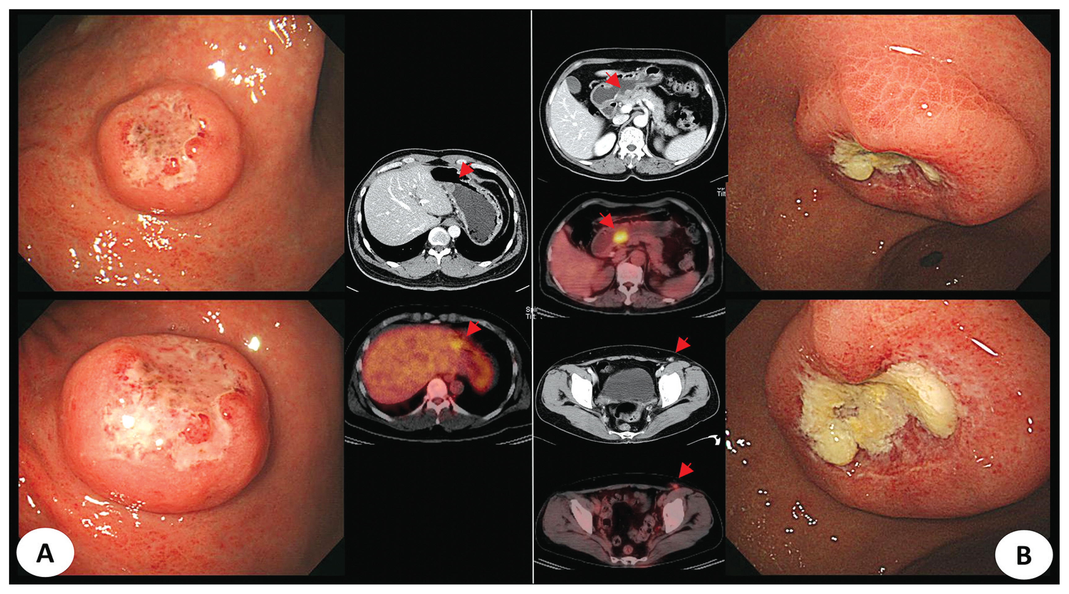
Fig. 3 PET-CT features of endoscopically polypoid mass-like lesions in primary DLBCL (A) and secondary DLBCL (B) cases (Abbreviation. PET-CT, positron emission tomography-computed tomography; DLBCL, diffuse large B-cell lymphoma).

Fig. 4 Endoscopically ulceroinfiltrative lesions of two cases. (A) Primary gastric lymphoma which was diagnosed as mantle-cell lymphoma. (B) Secondary gastric lymphoma which was diagnosed as DLBLC. (C) Multiple involved lymph nodes and organs were observed on PET-CT in same patients of B (Abbreviation. DLBCL, diffuse large B-cell lymphoma; PET-CT, positron emission tomography-computed tomography).

Fig. 5 Two cases of mixed type with two or more endoscopic findings. (A) Primary DLBLC. (B) Secondary DLBLC. (C) Hypermetabolism of stomach and multiple distant lymph node was seen on PET-CT in cases of B (Abbreviation. DLBCL, diffuse large B-cell lymphoma; PET-CT, positron emission tomography-computed tomography).
Reference
-
1. Ghimire P, Wu GY, Zhu L. Primary gastrointestinal lymphoma. World J Gastroenterol. 2011; 17:697–707.
Article2. Freeman C, Berg JW, Cutler SJ. Occurrence and prognosis of extranodal lymphomas. Cancer. 1972; 29:252–60.
Article3. Herrmann R, Panahon AM, Barcos MP, Walsh D, Stutzman L. Gastrointestinal involvement in non-Hodgkin’s lymphoma. Cancer. 1980; 46:215–22.
Article4. Andriani A, Zullo A, Di Raimondo F, Patti C, Tedeschi L, Recine U, et al. Clinical and endoscopic presentation of primary gastric lymphoma: a multicentre study. Aliment Pharmacol Ther. 2006; 23:721–6.
Article5. Doglioni C, Ponzoni M, Ferreri AJ, Savio A. Gastric lymphoma: the histology report. Dig Liver Dis. 2011; 43(Suppl 4):S310–8.
Article6. Ferreri AJ, Montalbán C. Primary diffuse large B-cell lymphoma of the stomach. Crit Rev Oncol Hematol. 2007; 63:65–71.
Article7. Dawson IM, Cornes JS, Morson BC. Primary malignant lymphoid tumours of the intestinal tract. Report of 37 cases with a study of factors influencing prognosis. Br J Surg. 1961; 49:80–9.
Article8. Palmer ED. The sarcomas of the stomach: a review with reference to gross pathology and gastroscopic manifestations. Am J Dig Dis. 1950; 17:186–95.
Article9. Seifert E, Schulte F, Weismuller J, de Mas CR, Stolte M. Endoscopic and bioptic diagnosis of malignant non-Hodgkin’s lymphoma of the stomach. Endoscopy. 1993; 25:497–501.
Article10. Kolve M, Fischbach W, Greiner A, Wilms K. Differences in endoscopic and clinicopathological features of primary and secondary gastric non-Hodgkin’s lymphoma. German Gastrointestinal Lymphoma Study Group. Gastrointest Endosc. 1999; 49:307–15.11. Cui X, Zhou T, Jiang D, Liu H, Wang J, Yuan S, et al. Clinical manifestations and endoscopic presentations of gastric lymphoma: a multicenter seven year retrospective survey. Rev Esp Enferm Dig. 2017; 109:566–71.
Article12. Ge Z, Liu Z, Hu X. Anatomic distribution, clinical features, and survival data of 87 cases primary gastrointestinal lymphoma. World J Surg Oncol. 2016; 14:85.
Article13. Zeggai S, Harir N, Tou A, Medjamia M, Guenaoui K. Gastrointestinal lymphoma in Western Algeria: pattern of distribution and histological subtypes (retrospective study). J Gastrointest Oncol. 2016; 7:1011–6.
Article14. Li DM, Jiang YP. Gastrointestinal involvement by mantle cell lymphoma observed by endoscopy: A case report. Medicine (Baltimore). 2017; 96:e6321.
