CD20 Positive T Cell Lymphoma Involvement of Skin
- Affiliations
-
- 1Department of Dermatology, College of Medicine, Dong-A University, Busan, Korea. khkim@dau.ac.kr
Abstract
- CD20 positive T cell lymphoma is a rare condition that is associated with the coexpressions of CD20 and T cell markers, such as, CD3, CD5, or UCHL-1. Positivity for CD20 in this tumor represents an aberrant immunophenotype, but the presence of monoclonal T cell receptor (TCR) gene rearrangements and negativity for immunoglobulin heavy chain gene rearrangement indicate that this tumor is a T cell lymphoma. The majority of cases of CD20 positive T cell lymphoma have been reported as immature peripheral T cell lymphoma not otherwise specified. However, we believe that this disease is likely to be re-listed as a new disease entity after its pathogenesis has been elucidated and more cases have been evaluated. Here, we present a case of peripheral T cell lymphoma coexpressing CD20 and T cell markers with a demonstrable TCR gene rearrangement, in a patient who had been misdiagnosed as having B cell type lymphoma 4 years previously. We hypothesize that in this case initially circulating normal CD20+ T cell subsets underwent neoplastic transformation and CD20 positive T cell lymphoma subsequently developed in the lymph node, and then recurred in the skin due to systemic disease or metastasized from the nodal disease.
MeSH Terms
Figure
-

Fig. 1 Variably sized multiple crusted erythematous masses on the face, neck (A & B), and Left forearm (C).

Fig. 2 Diffuse and pandermal lymphocytic infiltration without epidermal involvement (A: H&E, ×40). The round tumor cells were small to medium sized with vesicular nuclei and prominent nucleoli and had a relatively monotonous appearance (B: H&E, ×400).
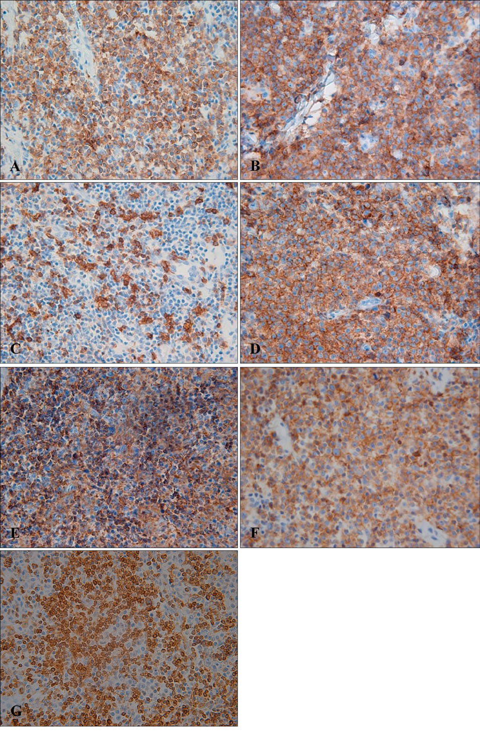
Fig. 3 Analysis of CD3 (A), CD4 (B), CD5 (C), UCHL-1 (D), CD20 (E), and for aCD79a (F) and bcl-2 (G) (immunoperoxidase stain, ×400).
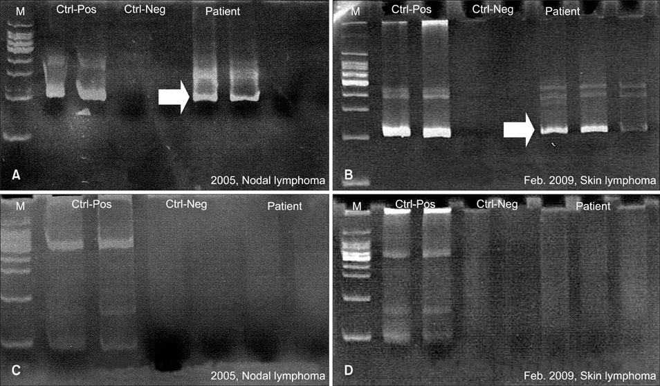
Fig. 4 T cell receptor gamma gene rearrangement showed monoclononality at around 200 bp in the present and previous biopsy specimens (A, B), but immunoglobulin heavy chain gene rearrangement showed no monoclonality in either specimen (C, D).
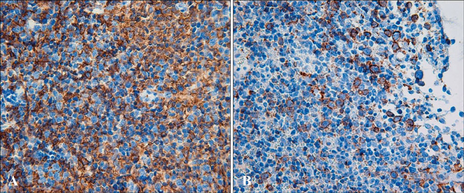
Fig. 5 The immunophenotypic evaluation performed on a neck lymph node excised at the biopsy performed in 2005, was strongly positive for CD20 (A, ×400) and weakly reactive with UCHL-1 (B, ×400) in the cytoplasm of atypical lymphocytes.

Fig. 6 Immunophenotypic evaluation from a neck lymph node excised in 2005 was positive for CD3 (A, ×400) and CD4 (B, ×400) and negative for CD30 (C, ×400).
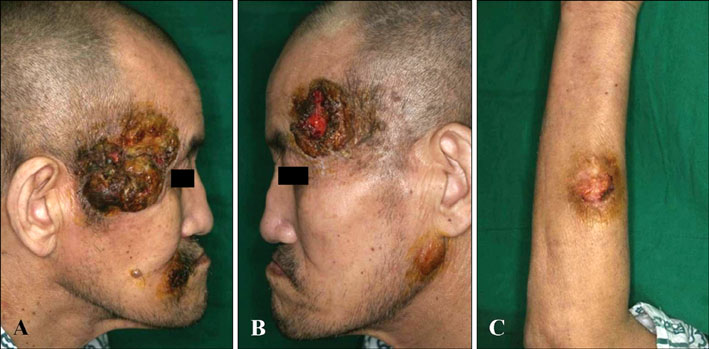
Fig. 7 The patient underwent ifosfamide, methotrexate, VP-16 (etoposide), and prednisolone chemotherapy and achieved partial remission with a decrease in mass size (A & B: face, neck, C: Left forearm).
Reference
-
1. Mohrmann RL, Arber DA. CD20-Positive peripheral T-cell lymphoma: report of a case after nodular sclerosis Hodgkin's disease and review of the literature. Mod Pathol. 2000. 13:1244–1252.
Article2. Balmer NN, Hughey L, Busam KJ, Reddy V, Andea AA. Primary cutaneous peripheral T-cell lymphoma with aberrant coexpression of CD20: case report and review of the literature. Am J Dermatopathol. 2009. 31:187–192.
Article3. Sen F, Kang S, Cangiarella J, Kamino H, Hymes K. CD20 positive mycosis fungoides: a case report. J Cutan Pathol. 2008. 35:398–403.
Article4. Magro CM, Seilstad KH, Porcu P, Morrison CD. Primary CD20+CD10+CD8+ T-cell lymphoma of the skin with IgH and TCR beta gene rearrangement. Am J Clin Pathol. 2006. 126:14–22.
Article5. Yokose N, Ogata K, Sugisaki Y, Mori S, Yamada T, An E, et al. CD20-positive T cell leukemia/lymphoma: case report and review of the literature. Ann Hematol. 2001. 80:372–375.
Article6. Quintanilla-Martinez L, Preffer F, Rubin D, Ferry JA, Harris NL. CD20+ T-cell lymphoma. Neoplastic transformation of a normal T-cell subset. Am J Clin Pathol. 1994. 102:483–489.
Article7. Warzynski MJ, Graham DM, Axtell RA, Zakem MH, Rotman RK. Low level CD20 expression on T cell malignancies. Cytometry. 1994. 18:88–92.
Article8. Buckner CL, Christiansen LR, Bourgeois D, Lazarchick JJ, Lazarchick J. CD20 positive T-cell lymphoma/leukemia: a rare entity with potential diagnostic pitfalls. Ann Clin Lab Sci. 2007. 37:263–267.9. Sun T, Akalin A, Rodacker M, Braun T. CD20 positive T cell lymphoma: is it a real entity? J Clin Pathol. 2004. 57:442–444.
Article10. Yao X, Teruya-Feldstein J, Raffeld M, Sorbara L, Jaffe ES. Peripheral T-cell lymphoma with aberrant expression of CD79a and CD20: a diagnostic pitfall. Mod Pathol. 2001. 14:105–110.
Article11. Rahemtullah A, Longtine JA, Harris NL, Dorn M, Zembowicz A, Quintanilla-Fend L, et al. CD20+ T-cell lymphoma: clinicopathologic analysis of 9 cases and a review of the literature. Am J Surg Pathol. 2008. 32:1593–1607.12. Balmer NN, Hughey L, Busam KJ, Reddy V, Andea AA. Primary cutaneous peripheral T-cell lymphoma with aberrant coexpression of CD20: case report and review of the literature. Am J Dermatopathol. 2009. 31:187–192.
Article13. Oshima H, Matsuzaki Y, Takeuchi S, Nakano H, Sawamura D. CD20+ primary cutaneous T-cell lymphoma presenting as a solitary extensive plaque. Br J Dermatol. 2009. 160:894–896.
Article14. Gill HS, Lau WH, Chan AC, Leung RY, Khong PL, Leung AY, et al. CD20 expression in natural killer T cell lymphoma. Histopathology. 2010. 57:157–159.
Article15. Xiao WB, Wang ZM, Wang LJ. CD20-positive T-cell lymphoma with indolent clinical behaviour. J Int Med Res. 2010. 38:1170–1174.
Article16. Tarkowski M. Expression and a role of CD30 in regulation of T-cell activity. Curr Opin Hematol. 2003. 10:267–271.
Article17. Murayama Y, Mukai R, Sata T, Matsunaga S, Noguchi A, Yoshikawa Y. Transient expression of CD20 antigen (pan B cell marker) in activated lymph node T cells. Microbiol Immunol. 1996. 40:467–471.
Article18. Venizelos ID, Tatsiou ZA, Mandala E. Primary cutaneous T-cell-rich B-cell lymphoma: a case report and literature review. Acta Dermatovenerol Alp Panonica Adriat. 2008. 17:177–181.19. Medeiros LJ, Elenitoba-Johnson KS. Anaplastic large cell lymphoma. Am J Clin Pathol. 2007. 127:707–722.
Article20. Cerny T, Borisch B, Introna M, Johnson P, Rose AL. Mechanism of action of rituximab. Anticancer Drugs. 2002. 13:Suppl 2. S3–S10.
Article21. Hamilton-Dutoit SJ, Pallesen G. B cell associated monoclonal antibody L26 may occasionally label T cell lymphomas. APMIS. 1989. 97:1033–1036.
Article22. Norton AJ, Isaacson PG. Monoclonal antibody L26: an antibody that is reactive with normal and neoplastic B lymphocytes in routinely fixed and paraffin wax embedded tissues. J Clin Pathol. 1987. 40:1405–1412.
Article
