First reported case of fetal aortic valvuloplasty in Asia
- Affiliations
-
- 1Department of Obstetrics and Gynecology, University of Ulsan College of Medicine, Asan Medical Center, Seoul, Korea. hswon@amc.seoul.kr
- 2Division of Neonatology, Department of Pediatrics, University of Ulsan College of Medicine, Asan Medical Center, Seoul, Korea.
- 3Department of Pediatric Cardiology, University of Ulsan College of Medicine, Asan Medical Center, Seoul, Korea.
- KMID: 2383216
- DOI: http://doi.org/10.5468/ogs.2017.60.1.106
Abstract
- Prenatal intervention of severe fetal aortic valve stenosis by ultrasound-guided percutaneous balloon valvuloplasty has been performed to prevent the progression to hypoplastic left heart syndrome, and achieve biventricular circulation in neonates. Here we report a case of fetal aortic valvuloplasty prenatally diagnosed with aortic stenosis at 24 weeks of gestation and showed worsening features on a follow-up echocardiography. Prenatal aortic valvuloplasty was performed at 29 weeks of gestation, and was a technical success. However, fetal bradycardia sustained, and an emergency cesarean delivery was performed. To the best of our knowledge, this is the first reported case of fetal aortic valvuloplasty which was performed in Asia.
MeSH Terms
Figure
-
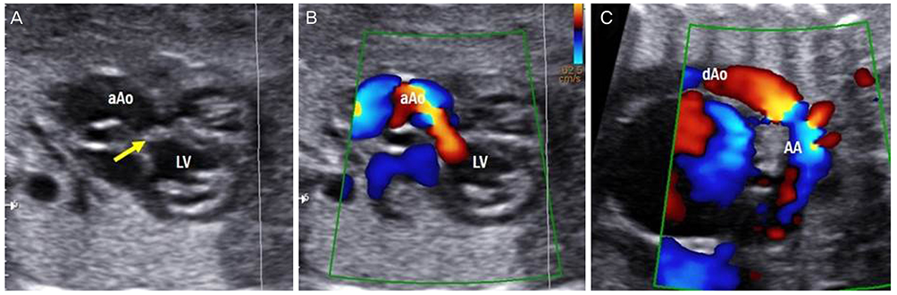
Fig. 1 The left ventricular outflow tract view of the heart at 24.1 weeks showing the thickening of the aortic valve (arrow in A), with turbulent flow across the aortic valve annulus on a color Doppler image (B). Follow-up evaluation at 26.1 weeks showing the retrograde flow in the aortic arch (C). aAo, ascending aorta; LV, left ventricle; dAo, descending aorta; AA, aortic arch.
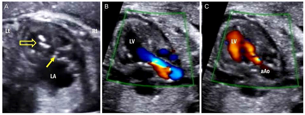
Fig. 2 The ultrasonographic findings reveal that the cannula is located in the LV (open arrow in A), and the thickened aortic valve is demonstrated (solid arrow in A). Immediately after the balloon dilation, the ultrasonographic findings demonstrate the increased forward flow across the aortic valve (B), and a newly developed aortic regurgitation by color Doppler (C). Lt, left; Rt, right; LA, left atrium; LV, left ventricle; aAo, ascending aorta.
Reference
-
1. Allan LD, Sharland G, Tynan MJ. The natural history of the hypoplastic left heart syndrome. Int J Cardiol. 1989; 25:341–343.2. Simpson JM, Sharland GK. Natural history and outcome of aortic stenosis diagnosed prenatally. Heart. 1997; 77:205–210.3. Maxwell D, Allan L, Tynan MJ. Balloon dilatation of the aortic valve in the fetus: a report of two cases. Br Heart J. 1991; 65:256–258.4. Freud LR, McElhinney DB, Marshall AC, Marx GR, Friedman KG, del Nido PJ, et al. Fetal aortic valvuloplasty for evolving hypoplastic left heart syndrome: postnatal outcomes of the first 100 patients. Circulation. 2014; 130:638–645.5. Moon-Grady AJ, Morris SA, Belfort M, Chmait R, Dangel J, Devlieger R, et al. International fetal cardiac intervention registry: a worldwide collaborative description and preliminary outcomes. J Am Coll Cardiol. 2015; 66:388–399.6. Tworetzky W, Wilkins-Haug L, Jennings RW, van der Velde ME, Marshall AC, Marx GR, et al. Balloon dilation of severe aortic stenosis in the fetus: potential for prevention of hypoplastic left heart syndrome. Candidate selection, technique, and results of successful intervention. Circulation. 2004; 110:2125–2131.7. Schidlow DN, Tworetzky W, Wilkins-Haug LE. Percutaneous fetal cardiac interventions for structural heart disease. Am J Perinatol. 2014; 31:629–636.8. Makikallio K, McElhinney DB, Levine JC, Marx GR, Colan SD, Marshall AC, et al. Fetal aortic valve stenosis and the evolution of hypoplastic left heart syndrome: patient selection for fetal intervention. Circulation. 2006; 113:1401–1405.9. Gardiner HM, Kovacevic A, Tulzer G, Sarkola T, Herberg U, Dangel J, et al. Natural history of 107 cases of fetal aortic stenosis from a European multicenter retrospective study. Ultrasound Obstet Gynecol. 2016; 48:373–381.10. Arzt W, Wertaschnigg D, Veit I, Klement F, Gitter R, Tulzer G. Intrauterine aortic valvuloplasty in fetuses with critical aortic stenosis: experience and results of 24 procedures. Ultrasound Obstet Gynecol. 2011; 37:689–695.11. McElhinney DB, Marshall AC, Wilkins-Haug LE, Brown DW, Benson CB, Silva V, et al. Predictors of technical success and postnatal biventricular outcome after in utero aortic valvuloplasty for aortic stenosis with evolving hypoplastic left heart syndrome. Circulation. 2009; 120:1482–1490.
- Full Text Links
-
- Actions
-
Cited
- CITED
-
- Close
- Share
-
- Similar articles
-
- Avulsion of Aortic Commissure: Rare Cause of Aortic Regurgitation: 2 case reports
- Percutaneous transluminal balloon valvuloplasty for congenital pulmonary valve stenosis and aortic valve stenosis
- The Management of Three Infants with Critical Valvular Aortic Stenosis by Transcarotid Balloon Aortic Valvuloplasty
- Clinical Experiences of Congenital Aortic Stenosis
- Percutaneous Balloon Valvuloplasty in Children with Pulmonary and Aortic Valvular Stenosis
