Gastric Langerhans Cell Histiocytosis: Case Report and Review of the Literature
- Affiliations
-
- 1Department of Pathology, Pusan National University Hospital, Pusan National University School of Medicine, Busan, Korea. pdy220@pusan.ac.kr
- 2Biomedical Research Institute, Pusan National University Hospital, Busan, Korea.
- KMID: 2381400
- DOI: http://doi.org/10.4132/jptm.2015.05.19
Abstract
- No abstract available.
MeSH Terms
Figure
-
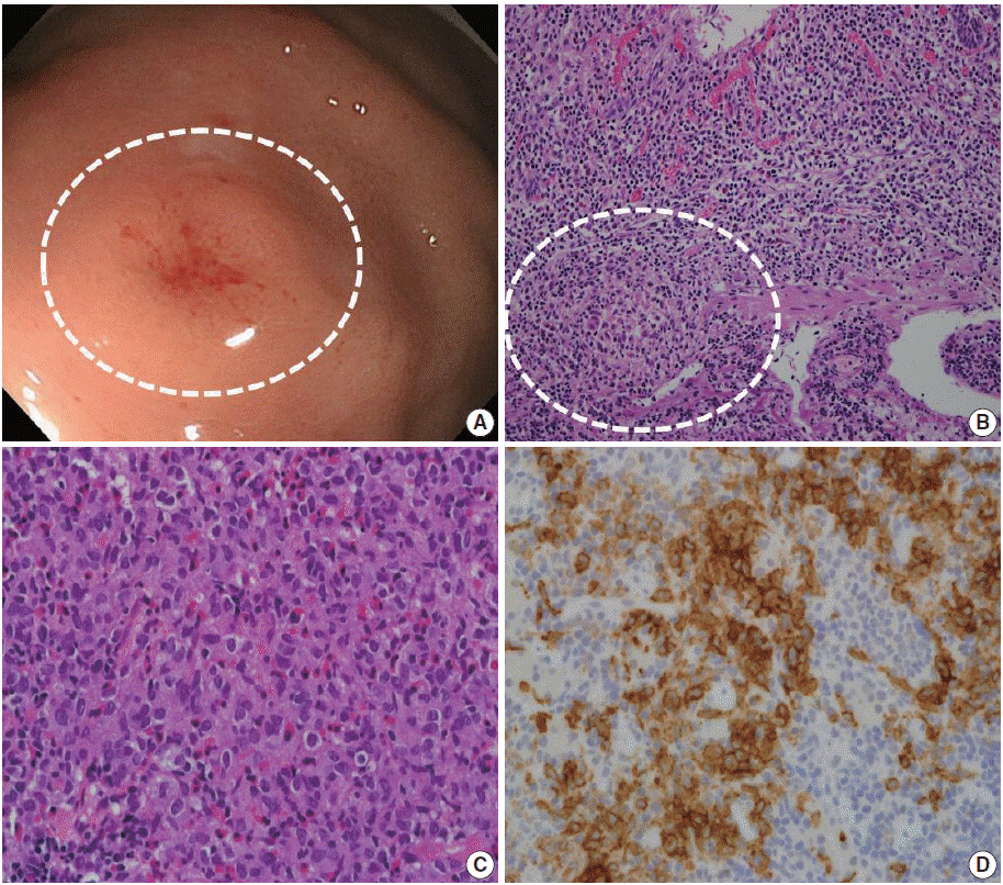
Fig. 1. Endoscopic and histologic finding of gastric Langerhans cell histiocytosis. (A) A mild elevated mucosal lesion (1 cm in size) with central erosion is observed upon gastroenteroscopy (circle). The lesion is located in the fundus of the stomach. (B) Microscopic analysis of the endoscopic submucosal dissection specimen. Focal histiocytic cell aggregates are present in the lamina propria and muscularis mucosa, with abundant eosinophils and other inflammatory cells. (C) Microscopic analysis of the endoscopic biopsy specimen reveals histiocytic cell aggregates in the lamina propria of the mucosa, with abundant eosinophil infiltration. Lymphocytes and plasma cells are also observed. The histiocytic cells show an irregular nuclear membrane and groove. These cells have abundant and granular eosinophilic to clear cytoplasm. (D) Immunohistochemistry for CD1a. The histiocytic cells show positive staining for CD1a.
Reference
-
1. Abla O, Egeler RM, Weitzman S. Langerhans cell histiocytosis: current concepts and treatments. Cancer Treat Rev. 2010; 36:354–9.
Article2. Behdad A, Owens SR. Langerhans cell histiocytosis involving the gastrointestinal tract. Arch Pathol Lab Med. 2014; 138:1350–2.
Article3. Detlefsen S, Fagerberg CR, Ousager LB, et al. Histiocytic disorders of the gastrointestinal tract. Hum Pathol. 2013; 44:683–96.
Article4. Singhi AD, Montgomery EA. Gastrointestinal tract langerhans cell histiocytosis: a clinicopathologic study of 12 patients. Am J Surg Pathol. 2011; 35:305–10.5. Vetter-Laracy S, Salinas JA, Martin-Santiago A, Guibelalde M, Balliu PR. Digestive tract symptoms in congenital langerhans cell histiocytosis: a fatal condition in an illness usually considered benign. J Pediatr Hematol Oncol. 2014; 36:426–9.6. Iwafuchi M, Watanabe H, Shiratsuka M. Primary benign histiocytosis X of the stomach: a report of a case showing spontaneous remission after 5 1/2 years. Am J Surg Pathol. 1990; 14:489–96.7. Nihei K, Terashima K, Aoyama K, Imai Y, Sato H. Benign histiocytosis X of stomach: previously undescribed lesion. Acta Pathol Jpn. 1983; 33:577–88.
Article8. Vazquez JJ, Ayestaran JR. Eosinophilic granuloma of the stomach similar to that of bone: light and electron microscopic study. Virchows Arch A Pathol Anat Histol. 1975; 366:107–11.9. Lee CK, Lee SH, Cho HD. Localized Langerhans cell histiocytosis of the stomach treated by endoscopic submucosal dissection. Endoscopy. 2011; 43 Suppl 2:E268–9.
Article10. Wada R, Yagihashi S, Konta R, Ueda T, Izumiyama T. Gastric polyposis caused by multifocal histiocytosis X. Gut. 1992; 33:994–6.
Article
- Full Text Links
-
- Actions
-
Cited
- CITED
-
- Close
- Share
-
- Similar articles
-
- A Case of Gastric Langerhans Cell Histiocytosis Showing Hypertrophic Folds
- Langerhans cell histiocytosis of the mandible: two case reports and literature review
- Spontaneous Pneumothorax due to Pulmonary Invasion in Multisystemic Langerhans Cell Histiocytosis: A case report
- A Case of Gastric Langerhans Cell Histiocytosis with Spontaneous Regression
- A Case of Secondary Sclerosing Cholangitis in Langerhans Cell Histiocytosis
