Acral Lentiginous Melanoma, Indolent Subtype Diagnosed by En Bloc Excision: A Case Report
- Affiliations
-
- 1Department of Dermatology, SMG-SNU Boramae Medical Center, Seoul, Korea. snuhdm@gmail.com
- 2Department of Pathology, SMG-SNU Boramae Medical Center, Seoul, Korea.
- KMID: 2378533
- DOI: http://doi.org/10.5021/ad.2017.29.3.327
Abstract
- Nail unit melanoma is a type of acral lentiginous melanoma and requires histopathologic examination for a confirmed diagnosis. However, inadequate biopsy techniques make definitive diagnosis difficult. A 61-year-old man presented with progressive nail pigmentation for 15 years, which was clinically highly suspicious for malignancy. Acral lentiginous melanoma was not detected in punch and longitudinal biopsy specimens, but en bloc excision tissue revealed melanoma. Acral lentiginous melanoma is known to have a heterogeneous pathologic manifestation depending on the pigmented region and the time it takes to progress. In this regard, en bloc excision can be considered as a first-line biopsy technique to diagnose acral lentiginous melanoma, indolent subtype.
Figure
-

Fig. 1 (A) Dark black colored pigmented band with about 1-cm width on the left thumbnail. Ulceration, onychodystrophy, and Hutchinson's sign were observed. (B) Left lateral portion of the nail unit was longitudinally biopsied, enclosing nail folds, hyponychium, and nail matrix.

Fig. 2 (A) Prominent pigmentation in the nail plate without atypical melanocytes (H&E; ×100, inset: ×50). (B) Bland pigmentation was in the basal layer of hyponycium (H&E; ×100, inset: ×10). (C) Significant pigmentation was observed in the nail bed without atypical melanocytes (H&E; ×400, inset: ×100).
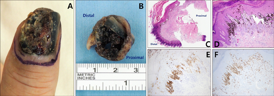
Fig. 3 (A) Two months later, the nail unit presented with plate destruction and intermittent bleeding from the nailbed. (B) En bloc excisional specimen of the nail unit. (C) Scattered pigmentation was observed in the hyponychium and the nail bed with lymphocytic infiltration in the onychodermis layer (H&E, ×10). (D) Atypical melanocytes with hyperchromatic nuclei invaded the dermis layer (H&E, ×100). (E, F) Immunohistochemical stains for human melanoma black 45 and Ki-67 were positive (×100).
Cited by 1 articles
-
Diagnosis and treatment of subungual melanoma
Joon Seok Oh, Byung Jun Kim
Arch Hand Microsurg. 2023;28(3):125-136. doi: 10.12790/ahm.22.0072.
Reference
-
1. Dawber RP, Colver GB. The spectrum of malignant melanoma of the nail apparatus. Semin Dermatol. 1991; 10:82–87.2. Ishihara K, Saida T, Yamamoto A. Japanese Skin Cancer Society Prognosis and Statistical Investigation Committee. Updated statistical data for malignant melanoma in Japan. Int J Clin Oncol. 2001; 6:109–116.
Article3. Kim JH, Park JH, Lee DY. Site distribution of cutaneous melanoma in South Korea: a retrospective study at a single tertiary institution. Int J Dermatol. 2015; 54:e38–e39.
Article4. Tan KB, Moncrieff M, Thompson JF, McCarthy SW, Shaw HM, Quinn MJ, et al. Subungual melanoma: a study of 124 cases highlighting features of early lesions, potential pitfalls in diagnosis, and guidelines for histologic reporting. Am J Surg Pathol. 2007; 31:1902–1912.5. Baran R, Kechijian P. Longitudinal melanonychia (melanonychia striata): diagnosis and management. J Am Acad Dermatol. 1989; 21:1165–1175.
Article6. Levit EK, Kagen MH, Scher RK, Grossman M, Altman E. The ABC rule for clinical detection of subungual melanoma. J Am Acad Dermatol. 2000; 42:269–274.7. Park HS, Cho KH. Acral lentiginous melanoma in situ: a diagnostic and management challenge. Cancers (Basel). 2010; 2:642–652.
Article8. Kim JY, Choi M, Jo SJ, Min HS, Cho KH. Acral lentiginous melanoma: indolent subtype with long radial growth phase. Am J Dermatopathol. 2014; 36:142–147.9. Lee DY. Variable sized cellular remnants in the nail plate of longitudinal melanonychia: evidence of subungual melanoma. Ann Dermatol. 2015; 27:328–329.
Article10. Phan A, Dalle S, Touzet S, Ronger-Savlé S, Balme B, Thomas L. Dermoscopic features of acral lentiginous melanoma in a large series of 110 cases in a white population. Br J Dermatol. 2010; 162:765–771.
Article11. Izumi M, Ohara K, Hoashi T, Nakayama H, Chiu CS, Nagai T, et al. Subungual melanoma: histological examination of 50 cases from early stage to bone invasion. J Dermatol. 2008; 35:695–703.
Article12. Shin HT, Park SW, Lee DY, Jang KT, Mun GH, Cheung L. A case of subungual melanoma with tumor invasion sparing the nail matrix dermis. Ann Dermatol. 2014; 26:655–657.
Article13. Shin HT, Jang KT, Mun GH, Lee DY, Lee JB. Histopathological analysis of the progression pattern of subungual melanoma: late tendency of dermal invasion in the nail matrix area. Mod Pathol. 2014; 27:1461–1467.
Article14. Blessing K, Sanders DS, Grant JJ. Comparison of immunohistochemical staining of the novel antibody melan-A with S100 protein and HMB-45 in malignant melanoma and melanoma variants. Histopathology. 1998; 32:139–146.
Article15. Sureda N, Phan A, Poulalhon N, Balme B, Dalle S, Thomas L. Conservative surgical management of subungual (matrix derived) melanoma: report of seven cases and literature review. Br J Dermatol. 2011; 165:852–858.
Article
- Full Text Links
-
- Actions
-
Cited
- CITED
-
- Close
- Share
-
- Similar articles
-
- A Case of Acral Lentiginous Melanoma
- Acral Lentiginous Melanoma in situ
- A Case of Thin Acral Lentiginous Melanoma with Lymph Node Metastasis and Regression
- Acral Lentiginous Melanoma Developing during Long-standing Atypical Melanosis: Usefulness of Dermoscopy for Detection of Early Acral Melanoma
- A Case of Acral Lentiginous Melanoma
