Acral Lentiginous Melanoma Developing during Long-standing Atypical Melanosis: Usefulness of Dermoscopy for Detection of Early Acral Melanoma
- Affiliations
-
- 1Department of Dermatology, Korea University College of Medicine, Seoul, Korea. kumcihk@korea.ac.kr
- KMID: 2171940
- DOI: http://doi.org/10.5021/ad.2011.23.3.400
Abstract
- Clinical guidelines suggest that suspicious pigmented lesions of the plantar or palmar area require biopsy for early detection of acral melanoma. We present here a case of acral lentiginous melanoma in which various melanocytic atypia was observed at each biopsy site, including focal melanocytic proliferation. We suggest that this atypical melanosis is part of a contiguous phase of invasive tumor growth, which is known as the very early stage of melanoma in situ. In addition, noninvasive dermoscopy has been effective for the early discovery of hidden lesions of acral melanoma.
Keyword
MeSH Terms
Figure
-

Fig. 1 A 1.5×0.8-cm hyperkeratotic patch with irregular border and variegated color on the left heel. The lesion was surrounded by a sharply demarcated brownish patch with some mottled dark pigmentation. Initial punch biopsy site (white arrow).
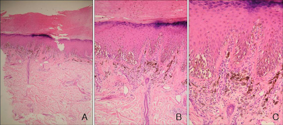
Fig. 2 Increase in basal melanocytes and hyperpigmentation with focally uniform, severe cytologic atypia of melanocytes (A: H&E, ×40, B: H&E, ×100, C: H&E, ×200).
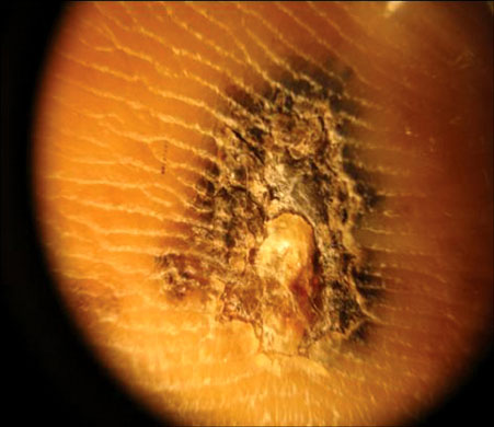
Fig. 3 Typical parallel ridge pattern and abrupt cut-off of pigmentation observed upon dermoscopy.
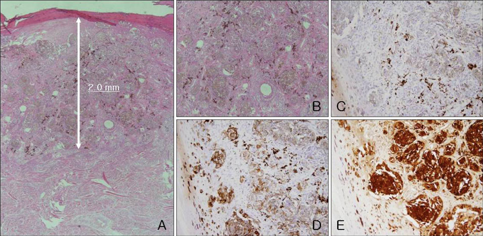
Fig. 4 (A, B) Atypical melanocytes are nested at the upper dermis, with a Breslow thickness of 2.0 mm (A: H&E, ×40, B: H&E, ×200), (C) Melan A(-) (H&E, ×200), (D) HMB45(+, focal) (H&E, ×200), (E) S-100(+) (H&E, ×200).

Fig. 5 Histologic findings of peripheral brownish patch area, melanocytic proliferation predominantly in the crista profunda intermedia and diffuse basal hyperpigmenation (A: H&E, ×40, B: H&E, ×100, C: H&E, ×200).
Reference
-
1. Chiu HH, Hu SC, Ke CL, Cheng ST. Dermoscopy identifies histopathologically indiscernible malignant lesion of atypical melanosis of the foot, an early lesion of acral lentiginous melanoma in situ. Dermatol Surg. 2008. 34:979–983.
Article2. Paek SC, Sober AJ, Tsao H, Mihm MC Jr, Johnson TM. Wolff K, Goldsmith LA, Katz SI, Gilchrest BA, Paller AS, Leffell DJ, editors. Cutaneous melanoma. Fitzpatrick's dermatology in general medicine. 2008. 7th ed. New York: McGraw-Hill;1134–1157.3. Metzger S, Ellwanger U, Stroebel W, Schiebel U, Rassner G, Fierlbeck G. Extent and consequences of physician delay in the diagnosis of acral melanoma. Melanoma Res. 1998. 8:181–186.
Article4. Somach SC, Taira JW, Pitha JV, Everett MA. Pigmented lesions in actinically damaged skin. Histopathologic comparison of biopsy and excisional specimens. Arch Dermatol. 1996. 132:1297–1302.
Article5. Mishima Y, Nakanishi T. Acral lentiginous melanoma and its precursor--heterogeneity of palmo-plantar melanomas. Pathology. 1985. 17:258–265.
Article6. Saida T. Malignant melanoma in situ on the sole of the foot. Its clinical and histopathologic characteristics. Am J Dermatopathol. 1989. 11:124–130.7. Nogita T, Wong TY, Ohara K, Mizushima J, Mihm MC Jr, Kawashima M. Atypical melanosis of the foot. A report of three cases in Japanese populations. Arch Dermatol. 1994. 130:1042–1045.
Article8. Cho KH, Kim BK, Lee DY, Minn KW. A case of acral melanocytic hyperplasia: a unique pigmented lesion mimicking acral lentiginous melanoma in situ. J Dermatol. 1996. 23:181–186.
Article9. Kwon IH, Lee JH, Cho KH. Acral lentiginous melanoma in situ: a study of nine cases. Am J Dermatopathol. 2004. 26:285–289.
Article10. Ishihara Y, Saida T, Miyazaki A, Koga H, Taniguchi A, Tsuchida T, et al. Early acral melanoma in situ: correlation between the parallel ridge pattern on dermoscopy and microscopic features. Am J Dermatopathol. 2006. 28:21–27.11. Braun RP, Rabinovitz HS, Oliviero M, Kopf AW, Saurat JH. Dermoscopy of pigmented skin lesions. J Am Acad Dermatol. 2005. 52:109–121.
Article12. Saida T, Miyazaki A, Oguchi S, Ishihara Y, Yamazaki Y, Murase S, et al. Significance of dermoscopic patterns in detecting malignant melanoma on acral volar skin: results of a multicenter study in Japan. Arch Dermatol. 2004. 140:1233–1238.13. Yamaura M, Takata M, Miyazaki A, Saida T. Specific dermoscopy patterns and amplifications of the cyclin D1 gene to define histopathologically unrecognizable early lesions of acral melanoma in situ. Arch Dermatol. 2005. 141:1413–1418.
Article14. Kilinc Karaarslan I, Akalin T, Unal I, Ozdemir F. Atypical melanosis of the foot showing a dermoscopic feature of the parallel ridge pattern. J Dermatol. 2007. 34:56–59.
Article
- Full Text Links
-
- Actions
-
Cited
- CITED
-
- Close
- Share
-
- Similar articles
-
- Acral Lentiginous Melanoma in situ
- A Case of Acral Lentiginous Melanoma
- Progression from Acral Lentiginous Melanoma in situ to Invasive Acral Lentiginous Melanoma
- Acral Lentiginous Melanoma, Indolent Subtype Diagnosed by En Bloc Excision: A Case Report
- A Case of Thin Acral Lentiginous Melanoma with Lymph Node Metastasis and Regression
