Clinical Analysis of Traumatic Cerebral Pseudoaneurysms
- Affiliations
-
- 1Department of Neurosurgery, Stroke Center, College of Medicine, Yonsei University, Severance Hospital, Seoul, Korea. leejw@yuhs.ac
- KMID: 2378270
- DOI: http://doi.org/10.13004/kjnt.2015.11.2.124
Abstract
OBJECTIVE
Traumatic pseudoaneurysms are rare but life-threatening lesions. We investigated the patients with these lesions to clarify their clinical characteristics and therapeutic strategies and we also reviewed the literatures on the treatment principles, possible options, and outcomes.
METHODS
There were a total of 8 patients who were treated with traumatic intracranial pseudoaneurysms between April 1980 and January 2009. Medical charts and the imaging studies were reviewed for analysis. The outcome was measured with modified Rankin Scale (mRS) score at 6 months after treatment.
RESULTS
All 8 patients were male and the mean age was 25 years old. Six of those were located at the cavernous segment of the internal carotid artery (ICA) and the other 2 was located at the M2 segment of middle cerebral artery. The causes of trauma were car accidents in 6, penetrating injury through the orbit in 1, and slip down injury in 1 patient. Massive epistaxis or hematemesis occurred in all patients with a pseudoaneurysm at the cavernous and ophthalmic segment of the ICA. All 6 patients of the cavernous and ophthalmic ICA group showed favorable outcome of mRS 0 to 1. The outcome of patients with middle cerebral artery pseudoaneurysm was mRS 2 to 3.
CONCLUSION
Upon prompt diagnosis and proper treatment planning, it is possible to achieve favorable outcome in these patients. Lesions located at the cavernous segment of the ICA favored endovascular treatment while those at the middle cerebral artery favored surgical treatment.
MeSH Terms
Figure
-
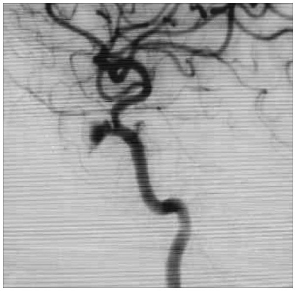
FIGURE 1 Lateral internal carotid artery (ICA) angiogram showing filling of a pseudosac at the cavernous segment of the ICA.
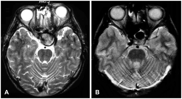
FIGURE 2 Magnetic resonance imaging showing a large pseudoaneurysm of cavernous segment of the internal carotid artery (A) and postoperative image showing decrease in aneurysm size (B).
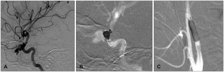
FIGURE 3 Lateral angiogram of the internal carotid artery (ICA) showing a pseudoaneurysm of the cavernous segment of the ICA (A). Coil embolization (B) and balloon occlusion (C) of the parent artery.
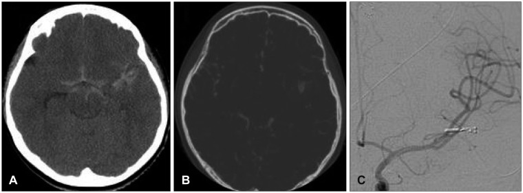
FIGURE 4 A: Non-contrast computed tomography (CT) showing subarachnoid hemorrhage. B: CT angiogram showing a pseudosac at the middle cerebral artery. C: Postoperative angiography showing no residual filling of the pseudosac.
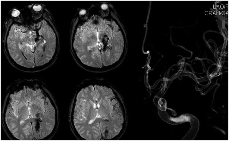
FIGURE 5 Gradient echo magnetic resonance imaging showing contusion injury of the brain and three-dimensional reconstructed view of a pseudoaneurysm of the middle cerebral artery.
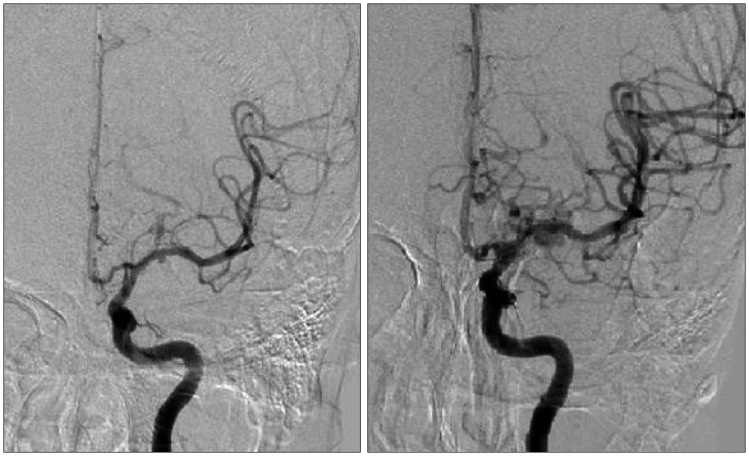
FIGURE 6 Initial and follow-up angiography of a middle cerebral artery pseudoaneurysm showing increase in size.
Cited by 1 articles
-
Internal Carotid Artery Pseudoaneurysm in a Patient Presenting With Recurrent Epistaxis: A Case Report and Literature Review
Hyejeen Kim, Ji Yun Choi
J Rhinol. 2024;31(1):46-51. doi: 10.18787/jr.2023.00072.
Reference
-
1. Adeel M, Ikram M. Post-traumatic pseudoaneurysm of internal carotid artery: a cause of intractable epistaxis. BMJ Case Rep. 2012; 5. 23. 2012:[Epub]. DOI: 10.1136/bcr.02.2012.5927.2. Amenta PS, Starke RM, Jabbour PM, Tjoumakaris SI, Gonzalez LF, Rosenwasser RH, et al. Successful treatment of a traumatic carotid pseudoaneurysm with the pipeline stent: case report and review of the literature. Surg Neurol Int. 2012; 3:160. PMID: 23372976.
Article3. Bavinzski G, Killer M, Knosp E, Ferraz-Leite H, Gruber A, Richling B. False aneurysms of the intracavernous carotid artery--report of 7 cases. Acta Neurochir (Wien). 1997; 139:37–43. PMID: 9059710.4. Cohen JE, Gomori JM, Segal R, Spivak A, Margolin E, Sviri G, et al. Results of endovascular treatment of traumatic intracranial aneurysms. Neurosurgery. 2008; 63:476–485. discussion 485-486PMID: 18812959.
Article5. Giorgianni A, Pellegrino C, Minotto R, Mercuri A, Frattini L, Baruzzi F, et al. Flow-diverter stenting of post-traumatic bilateral anterior cerebral artery pseudoaneurysm: a case report. Interv Neuroradiol. 2015; 21:23–28. PMID: 25934771.
Article6. Honeybul S, Barker S, Poitelea C, Ditchfield A. Multiple intracranial aneurysms presenting with epistaxis. J Clin Neurosci. 2006; 13:394–397. PMID: 16540324.
Article7. Horiuchi T, Nakagawa F, Miyatake M, Iwashita T, Tanaka Y, Hongo K. Traumatic middle cerebral artery aneurysm: case report and review of the literature. Neurosurg Rev. 2007; 30:263–267. discussion 267PMID: 17440757.
Article8. Larson PS, Reisner A, Morassutti DJ, Abdulhadi B, Harpring JE. Traumatic intracranial aneurysms. Neurosurg Focus. 2000; 8:e4. PMID: 16906700.
Article9. Lim YC, Kang JK, Chung J. Reconstructive stent-buttressed coil embolization of a traumatic pseudoaneurysm of the supraclinoid internal carotid artery. Acta Neurochir (Wien). 2012; 154:477–480. PMID: 22187050.
Article10. Maldonado-Naranjo A, Kshettry VR, Toth G, Bain M. Non-traumatic superior hypophyseal aneurysm with associated pseudoaneurysm presenting with massive epistaxis. Clin Neurol Neurosurg. 2013; 115:2251–2253. PMID: 23932468.
Article11. Nathoo N, Nadvi SS. Traumatic intracranial aneurysms following penetrating stab wounds to the head: two unusual cases and review of the literature. Cent Afr J Med. 1999; 45:213–217. PMID: 10697918.
Article12. Ohta M, Matsuno H. Proximal M2 false aneurysm after head trauma--case report. Neurol Med Chir (Tokyo). 2001; 41:131–134. PMID: 11372556.13. Ruiz-Juretschke F, Castro E, Mateo Sierra O, Iza B, Manuel Garbizu J, Fortea F, et al. Massive epistaxis resulting from an intracavernous internal carotid artery traumatic pseudoaneurysm: complete resolution with overlapping uncovered stents. Acta Neurochir (Wien). 2009; 151:1681–1684. PMID: 19350203.
Article14. Santos G, Lima T, Pereira S, Machado E. Traumatic middle cerebral artery aneurysm secondary to a gunshot wound. J Neuroimaging. 2013; 23:115–117. PMID: 21223435.
Article15. Tuchman A, Khalessi AA, Attenello FJ, Amar AP, Zada G. Delayed cavernous carotid artery pseudoaneurysm caused by absorbable plate following transsphenoidal surgery: case report and review of the literature. J Neurol Surg Rep. 2013; 74:10–16. PMID: 23943714.
Article
- Full Text Links
-
- Actions
-
Cited
- CITED
-
- Close
- Share
-
- Similar articles
-
- Traumatic middle meningeal artery pseudoaneurysm: Case report and review of literature
- Short-Term Clinical and Angiographic Outcome in Child with Traumatic Pseudoaneurysm in A2 Segment of Anterior Cerebral Artery after Endovascular Treatment: Case Report
- Angiographically Progressive Change of Traumatic Pseudoaneurysm Arising from the Middle Meningeal Artery
- Traumatic Aneurysm of Peripheral Cerebral Artery: A Case Report
- Post-Traumatic Cerebral Infarction
