J Gastric Cancer.
2011 Dec;11(4):195-199.
Clinicopathologic Significance of Gastric Adenocarcinoma with Neuroendocrine Features
- Affiliations
-
- 1Department of Surgery, National Police Hospital, Seoul, Korea.
- 2Department of Surgery, Ajou University School of Medicine, Suwon, Korea. hansu@ajou.ac.kr
Abstract
- PURPOSE
Composite neuroendocrine-exocrine carcinomas are malignancies that have two distinct components residing within the same tumor: an adenocarcinomatous portion and a neuroendocrine portion. This is rare in gastric cancers; however, poorly differentiated adenocarcinomas can sometimes reveal evidence of neuroendocrine features (NEF) or be 'mixed endocrine and exocrine carcinomas'. This study aimed to review NEF in gastric adenocarcinoma and to evaluate its prognostic significance.
MATERIALS AND METHODS
We selected 29 patients who were diagnosed with gastric adenocarcinoma with NEF and received gastrectomies at the Department of Surgery, Ajou University Hospital between January 2001 and December 2009. We analyzed the clinicopathologic features of gastric cancer with NEF and the prognosis associated with such tumors.
RESULTS
The pathologic result with respect to TNM staging of the gastric cancers with NEF were as follows: 5 cases of T1, 5 cases of T2, 10 cases of T3, and 9 cases of T4. There were 7 cases of N0, 7 cases of N1, 8 cases of N2 and 7 cases of N3. The staging of patients with NEF was higher than that of patients without NEF. Especially tumor lymphovascular invasion rate was 82.8%. The overall survival of patients with gastric cancer characterized by NEF was 73.8 months.
CONCLUSIONS
Positive NEF status might be correlated with clinicopathologic parameters such as a high stage and high frequency of regional lymph node metastasis.
MeSH Terms
Figure
-

Fig. 1 Histological finding of H&E stain and immunohistochemistry. (A) Adenocarcinoma with neuroendocrine features (H&E stain, ×200). (B) Positive in area of neuroendocrine tumor (Gold color) (Synaptophysin stain, ×200). (C) Positive in area of neuroendocrine tumor (Gold color) (Chromogranin A stain, ×200).
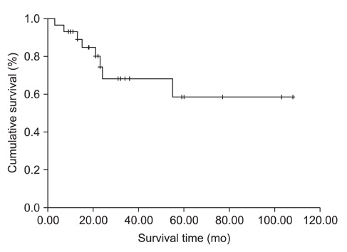
Fig. 2 Kaplan-Meier survival curves of gastric adenocarcinoma with neuroendocrine features.
Reference
-
1. Volante M, Righi L, Asioli S, Bussolati G, Papotti M. Goblet cell carcinoids and other mixed neuroendocrine/nonneuroendocrine neoplasms. Virchows Arch. 2007. 451:Suppl 1. S61–S69.
Article2. Rindi G, Arnold R, Bosman FT. Bosman FT, Carneiro F, Hruban RH, Theise ND, editors. Nomenclature and classification of neuroendocrine neoplasms of the digestive system. WHO classification of tumors of the digestive system. 2010. Lyon: IARC.
Article3. Lewin K. Carcinoid tumors and the mixed (composite) glandular-endocrine cell carcinomas. Am J Surg Pathol. 1987. 11:Suppl 1. 71–86.
Article4. Ooi A, Hayashi H, Katsuda S, Nakanishi I. Gastric carcinoma cells with endocrine differentiation show no evidence of proliferation. Hum Pathol. 1992. 23:736–741.
Article5. Park JM, Jang YJ, Kim JH, Park SS, Park SH, Kim SJ, et al. Gastric cancer histology: clinicopathologic characteristics and prognostic value. J Surg Oncol. 2008. 98:520–525.
Article6. Wanebo HJ, Kennedy BJ, Chmiel J, Steele G Jr, Winchester D, Osteen R. Cancer of the stomach. A patient care study by the American College of Surgeons. Ann Surg. 1993. 218:583–592.
Article7. Sim YK, Kim CY, Jeong YJ, Kim JH, Hwang Y, Yang DH. Changes of the clinicopathological characteristics and survival rates of gastric cancer with gastrectomy: 1990s vs early 2000s. J Korean Gastric Cancer Assoc. 2009. 9:200–206.
Article8. Seo WH, Seo BJ, Yu HJ, Lee WY, Lee HK. Analysis of prognostic factors in 1,435 surgically treated patients with gastric cancer. J Korean Gastric Cancer Assoc. 2009. 9:143–151.
Article9. Jeong O, Park YK. Clinicopathological features and surgical treatment of gastric cancer in South Korea: the results of 2009 nationwide survey on surgically treated gastric cancer patients. J Gastric Cancer. 2011. 11:69–77.
Article10. Kim KM, Kim MJ, Cho BK, Choi SW, Rhyu MG. Genetic evidence for the multi-step progression of mixed glandular-neuroendocrine gastric carcinomas. Virchows Arch. 2002. 440:85–93.
Article11. Eren F, Celikel C, Güllüoğlu B. Neuroendocrine differentiation in gastric adenocarcinomas; correlation with tumor stage and expression of VEGF and p53. Pathol Oncol Res. 2004. 10:47–51.12. Tamura T, Jobo T, Watanabe J, Kanai T, Kuramoto H. Neuroendocrine features in poorly differentiated endometrioid adenocarcinomas of the endometrium. Int J Gynecol Cancer. 2006. 16:821–826.13. Allen FJ, Van Velden DJ, Heyns CF. Are neuroendocrine cells of practical value as an independent prognostic parameter in prostate cancer? Br J Urol. 1995. 75:751–754.14. Wilson BS, Lloyd RV. Detection of chromogranin in neuroendocrine cells with a monoclonal antibody. Am J Pathol. 1984. 115:458–468.15. Gould VE, Wiedenmann B, Lee I, Schwechheimer K, Dockhorn-Dworniczak B, Radosevich JA, et al. Synaptophysin expression in neuroendocrine neoplasms as determined by immunocytochemistry. Am J Pathol. 1987. 126:243–257.
Article16. Waldum HL, Aase S, Kvetnoi I, Brenna E, Sandvik AK, Syversen U, et al. Neuroendocrine differentiation in human gastric carcinoma. Cancer. 1998. 83:435–444.
Article
- Full Text Links
-
- Actions
-
Cited
- CITED
-
- Close
- Share
-
- Similar articles
-
- A Case of a Gastric Composite Tumor with an Adenocarcinoma and a Large Cell Neuroendocrine Carcinoma
- Gastric Collision Tumor (Adenocarcinoma and Neuro-endocrine Carcinoma): A Report of Two Cases
- Metastatic Large Cell Neuroendocrine Carcinoma Combined with Gastric Adenocarcinoma
- A Case of Gastric Mixed Neuroendocrine-Nonneuroendocrine Neoplasm Composed With Adenoma and Neuroendocrine Tumor
- Gastric Adenocarcinoma with Coexistent Hepatoid Adenocarcinoma and Neuroendocrine Carcinoma: A Case Report
