Clinical Outcomes of an Optimized Prolate Ablation Procedure for Correcting Residual Refractive Errors Following Laser Surgery
- Affiliations
-
- 1The Institute of Vision Research, Department of Ophthalmology, Yonsei University College of Medicine, Seoul, Korea. tikim@yuhs.ac
- 2Lee Eye Clinic, Busan, Korea.
- 3Corneal Dystrophy Research Institute, Severance Biomedical Science Institute, and Brain Korea 21 Project for Medical Science, Department of Ophthalmology, Yonsei University College of Medicine, Seoul, Korea.
- KMID: 2368674
- DOI: http://doi.org/10.3341/kjo.2017.31.1.16
Abstract
- PURPOSE
The purpose of this study was to investigate the clinical efficacy of an optimized prolate ablation procedure for correcting residual refractive errors following laser surgery.
METHODS
We analyzed 24 eyes of 15 patients who underwent an optimized prolate ablation procedure for the correction of residual refractive errors following laser in situ keratomileusis, laser-assisted subepithelial keratectomy, or photorefractive keratectomy surgeries. Preoperative ophthalmic examinations were performed, and uncorrected distance visual acuity, corrected distance visual acuity, manifest refraction values (sphere, cylinder, and spherical equivalent), point spread function, modulation transfer function, corneal asphericity (Q value), ocular aberrations, and corneal haze measurements were obtained postoperatively at 1, 3, and 6 months.
RESULTS
Uncorrected distance visual acuity improved and refractive errors decreased significantly at 1, 3, and 6 months postoperatively. Total coma aberration increased at 3 and 6 months postoperatively, while changes in all other aberrations were not statistically significant. Similarly, no significant changes in point spread function were detected, but modulation transfer function increased significantly at the postoperative time points measured.
CONCLUSIONS
The optimized prolate ablation procedure was effective in terms of improving visual acuity and objective visual performance for the correction of persistent refractive errors following laser surgery.
MeSH Terms
Figure
-
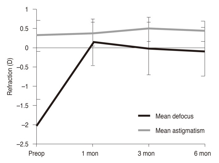
Fig. 1 Postoperative stability of refractive errors. D = diopters; Preop = preoperative.
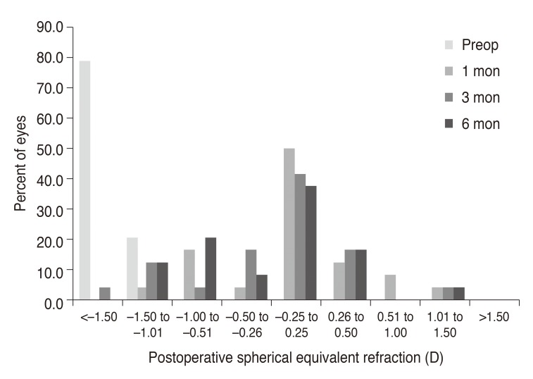
Fig. 2 Postoperative changes in spherical equivalent of manifest refraction. Preop = preoperative; D = diopters.
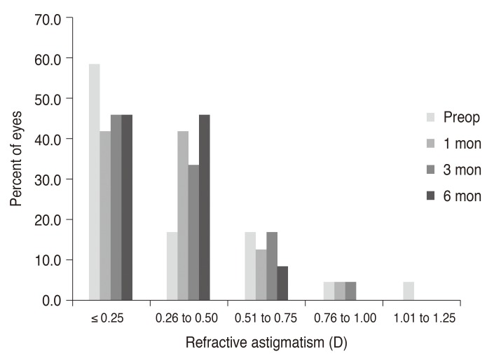
Fig. 3 Postoperative changes in spherical equivalent of manifest refraction. Preop = preoperative; D = diopters.
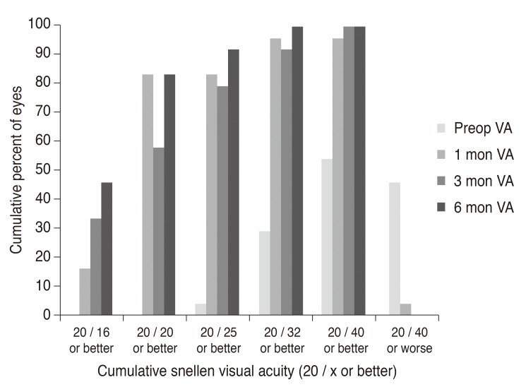
Fig. 4 Postoperative changes in astigmatic error of manifest refraction. Preop = preoperative; D = diopters.
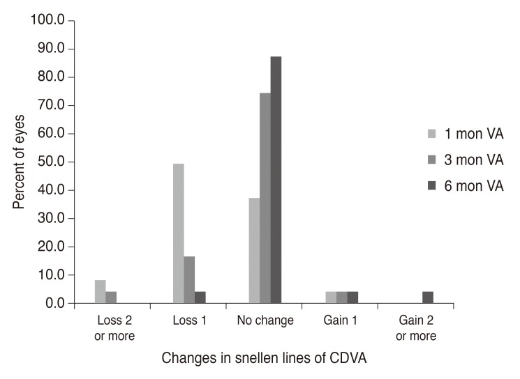
Fig. 5 Postoperative distribution of uncorrected visual acuity. Preop = preoperative; VA = visual acuity.
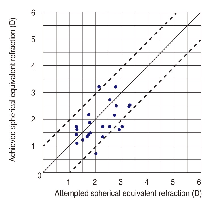
Fig. 6 Attempted versus achieved postoperative spherical equivalent. D = diopters.
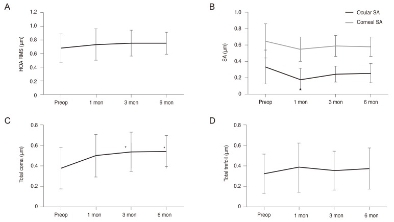
Fig. 7 Postoperative changes in ocular aberrations. (A) Total ocular HOA RMS, (B) ocular and corneal spherical aberration, (C) total coma aberrations, (D) total trefoil aberrations. HOA = higher-order aberration; RMS = root mean square; Preop = preoperative; SA = spherical aberration. *p < 0.05. All aberrations are reported for a 6.0-mm pupil plotted to the eighth Zernike order. Root-mean-square values are reported.
Reference
-
1. Rashad KM. Laser in situ keratomileusis retreatment for residual myopia and astigmatism. J Refract Surg. 2000; 16:170–176. PMID: 10766386.
Article2. Hersh PS, Fry KL, Bishop DS. Incidence and associations of retreatment after LASIK. Ophthalmology. 2003; 110:748–754. PMID: 12689897.
Article3. Alio JL, Muftuoglu O, Ortiz D, et al. Ten-year follow-up of laser in situ keratomileusis for high myopia. Am J Ophthalmol. 2008; 145:55–64. PMID: 17996210.4. Kanellopoulos AJ, Asimellis G. Refractive and keratometric stability in high myopic LASIK with high-frequency femtosecond and excimer lasers. J Refract Surg. 2013; 29:832–837. PMID: 24088061.
Article5. Aslanides IM, Kolli S, Padroni S, Arba Mosquera S. Stability of therapeutic retreatment of corneal wavefront customized ablation with the SCHWIND CAM: 4-year data. J Refract Surg. 2012; 28:347–352. PMID: 22515177.
Article6. Bragheeth MA, Fares U, Dua HS. Re-treatment after laser in situ keratomileusis for correction of myopia and myopic astigmatism. Br J Ophthalmol. 2008; 92:1506–1510. PMID: 18757469.
Article7. Toda I, Yamamoto T, Ito M, et al. Topography-guided ablation for treatment of patients with irregular astigmatism. J Refract Surg. 2007; 23:118–125. PMID: 17326350.
Article8. El-Danasoury A, Bains HS. Optimized prolate corneal ablation: case report of the first treated eye. J Refract Surg. 2005; 21(5 Suppl):S598–S602. PMID: 16212286.
Article9. El Danasoury AM, Holladay J, Waring GO 3rd, et al. A contralateral, randomized comparison of optimized prolate ablation and conventional LASIK for myopia with the NIDEK excimer laser platform. J Refract Surg. 2012; 28:453–461. PMID: 22767164.
Article10. Fantes FE, Hanna KD, Waring GO 3rd, et al. Wound healing after excimer laser keratomileusis (photorefractive keratectomy) in monkeys. Arch Ophthalmol. 1990; 108:665–675. PMID: 2334323.
Article11. Maldonado MJ. Intraoperative MMC after excimer laser surgery for myopia. Ophthalmology. 2002; 109:826. PMID: 11986073.
Article12. Jain S, McCally RL, Connolly PJ, Azar DT. Mitomycin C reduces corneal light scattering after excimer keratectomy. Cornea. 2001; 20:45–49. PMID: 11189003.
Article13. Kang EC, Choi BJ, Kim EK, Kim TI. Clinical outcomes of optimized prolate ablation and custom aspheric treatment in laser-assisted subepithelial keratectomy. J Cataract Refract Surg. 2012; 38:445–452. PMID: 22340605.
Article14. Choi BJ, Park YM, Lee JS. Clinical outcomes between optical path difference custom aspheric treatment and optimized prolate ablation photorefractive keratectomy in myopia exceeding 8 diopters. Eye (Lond). 2015; 29:356–362. PMID: 25397788.
Article15. Chayet A, Bains HS. Prospective, randomized, double-blind, contralateral eye comparison of myopic LASIK with optimized aspheric or prolate ablations. J Refract Surg. 2012; 28:112–119. PMID: 22201324.
Article16. Alio JL, Pinero DP, Plaza Puche AB. Corneal wavefront-guided enhancement for high levels of corneal coma aberration after laser in situ keratomileusis. J Cataract Refract Surg. 2008; 34:222–231. PMID: 18242444.17. Kermani O, Schmiedt K, Oberheide U, Gerten G. Topographic- and wavefront-guided customized ablations with the NIDEK-EC5000CXII in LASIK for myopia. J Refract Surg. 2006; 22:754–763. PMID: 17061712.
Article18. Vinciguerra P, Azzolini M, Radice P, et al. A method for examining surface and interface irregularities after photorefractive keratectomy and laser in situ keratomileusis: predictor of optical and functional outcomes. J Refract Surg. 1998; 14(2 Suppl):S204–S206. PMID: 9571554.
Article19. Balestrazzi E, De Molfetta V, Spadea L, et al. Histological, immunohistochemical, and ultrastructural findings in human corneas after photorefractive keratectomy. J Refract Surg. 1995; 11:181–187. PMID: 7553088.20. Oshika T, Klyce SD, Applegate RA, et al. Comparison of corneal wavefront aberrations after photorefractive keratectomy and laser in situ keratomileusis. Am J Ophthalmol. 1999; 127:1–7. PMID: 9932992.
Article21. Vinciguerra P, Azzolini M, Airaghi P, et al. Effect of decreasing surface and interface irregularities after photorefractive keratectomy and laser in situ keratomileusis on optical and functional outcomes. J Refract Surg. 1998; 14(2 Suppl):S199–S203. PMID: 9571553.
Article22. Vinciguerra P, Camesasca FI, Randazzo A. One-year results of butterfly laser epithelial keratomileusis. J Refract Surg. 2003; 19(2 Suppl):S223–S226. PMID: 12699177.
Article23. Serrao S, Lombardo M. One-year results of photorefractive keratectomy with and without surface smoothing using the technolas 217C laser. J Refract Surg. 2004; 20:444–449. PMID: 15523955.
Article24. Bas AM, Onnis R. Excimer laser in situ keratomileusis for myopia. J Refract Surg. 1995; 11(3 Suppl):S229–S233. PMID: 7553096.
Article25. Mohan RR, Hutcheon AE, Choi R, et al. Apoptosis, necrosis, proliferation, and myofibroblast generation in the stroma following LASIK and PRK. Exp Eye Res. 2003; 76:71–87. PMID: 12589777.
Article26. Roberts C. Biomechanics of the cornea and wavefront-guided laser refractive surgery. J Refract Surg. 2002; 18:S589–S592. PMID: 12361163.
Article27. Lindsay R, Smith G, Atchison D. Descriptors of corneal shape. Optom Vis Sci. 1998; 75:156–158. PMID: 9503441.
Article28. Gatinel D, Haouat M, Hoang-Xuan T. A review of mathematical descriptors of corneal asphericity. J Fr Ophtalmol. 2002; 25:81–90. PMID: 11965125.
- Full Text Links
-
- Actions
-
Cited
- CITED
-
- Close
- Share
-
- Similar articles
-
- Clinical Outcomes of Combined Procedure of Astigmatic Keratotomy and Laser in situ Keratomileusis
- Management of Three Cases of Decentration after Laser In Situ Keratomileusis
- The Effects of Laser Refractive Surgery for Correcting Residual Refractive Error after Implantation of ReSTOR(R) Multifocal IOL
- Clinical Characteristics of Intermittent Exotropia Patients who Have Improved due to Corrected Refractive Errors
- Mechanical Method Versus Laser Method for the Corneal Epithelium Ablation in Excimer Laser Photorefractive Keratectomy
