J Korean Ophthalmol Soc.
2016 Mar;57(3):353-360. 10.3341/jkos.2016.57.3.353.
Clinical Outcomes of Combined Procedure of Astigmatic Keratotomy and Laser in situ Keratomileusis
- Affiliations
-
- 1Onnuri Smile Eye Clinic, Seoul, Korea. ytchungc@hanmail.net
- KMID: 2213235
- DOI: http://doi.org/10.3341/jkos.2016.57.3.353
Abstract
- PURPOSE
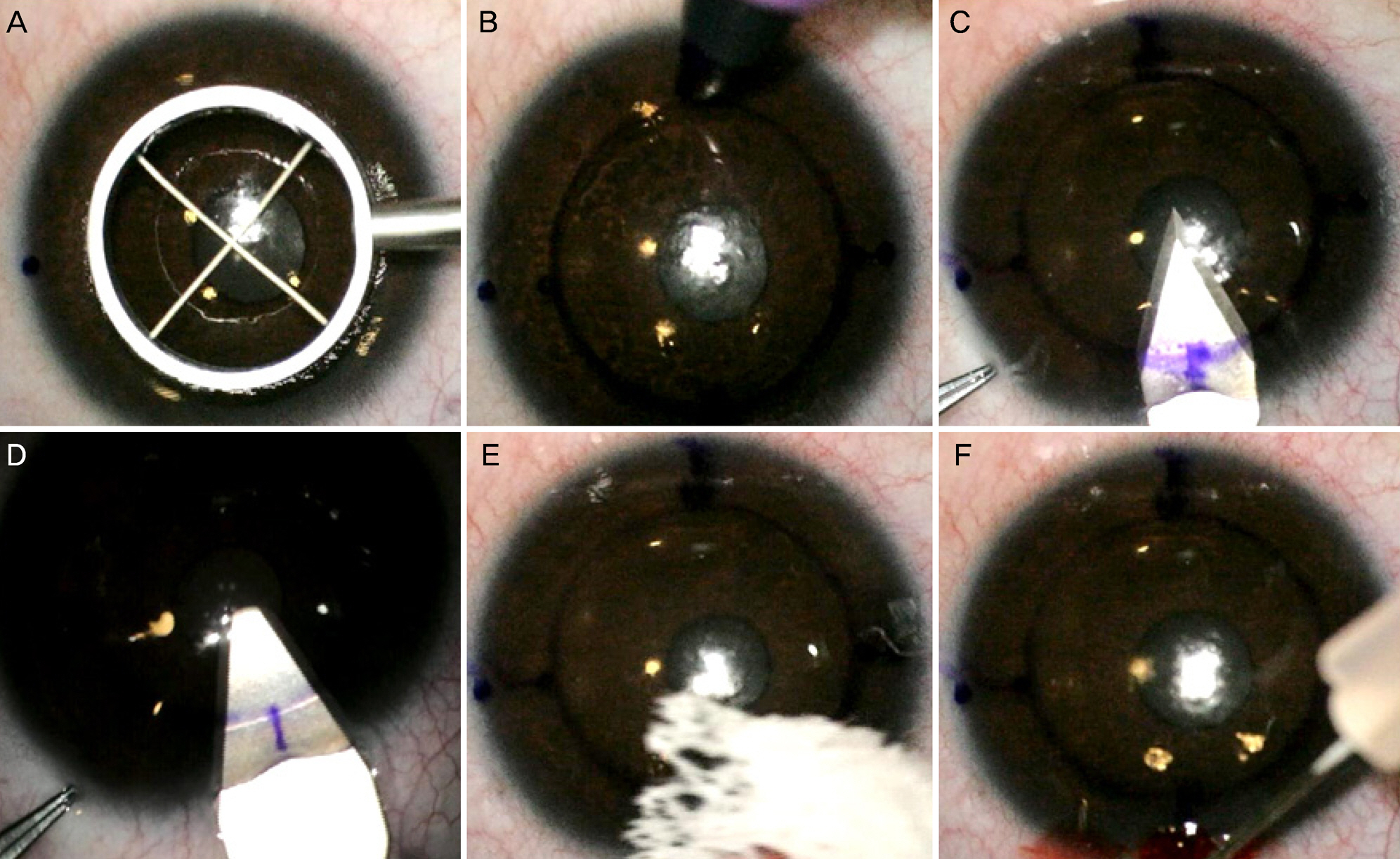
To evaluate the clinical outcomes of a combined procedure of astigmatic keratotomy (AK) and laser in situ keratomileusis (LASIK) for the correction of high astigmatism.
METHODS
Thirty-five eyes of 19 patients who had astigmatic keratotomy were studied. The patients had a secondary procedure, LASIK, to correct the residual refractive error. Follow-up visits were at 1 week, 1 month, 3 months, and 6 months. The outcome measures included uncorrected distance visual acuity, refractive error, efficacy, safety, and predictability. We compared preoperative and post-AK expected corneal ablation depth using an Amaris Ablation depth table.
RESULTS
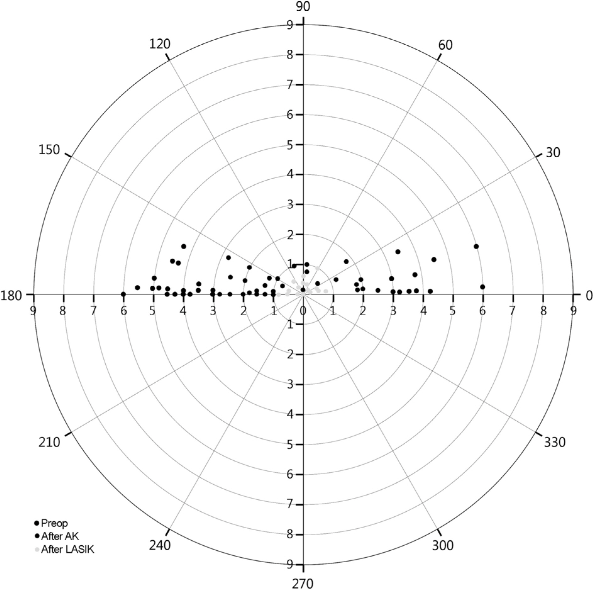
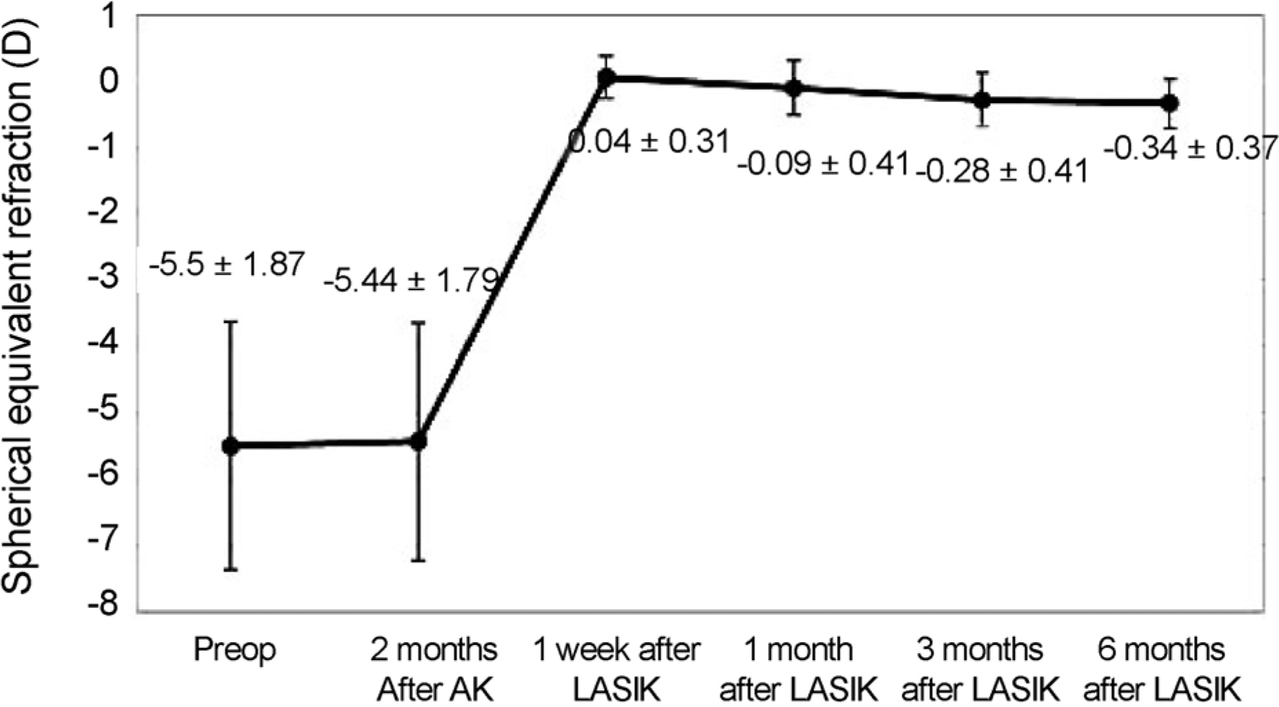
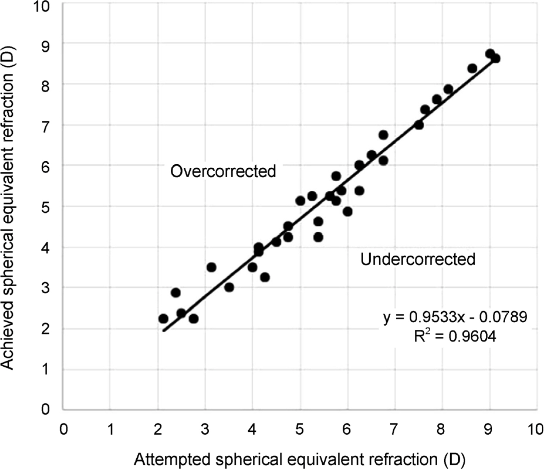
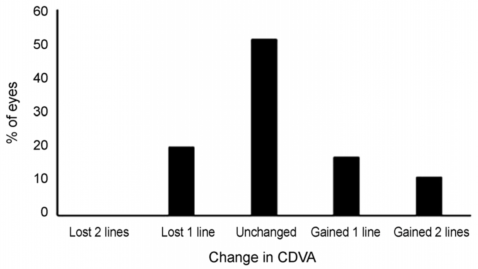
After astigmatic keratotomy, astigmatism was reduced by 61.43 ± 14.62%, and after LASIK, astigmatism was reduced by 91.65 ± 8.68%. Expected corneal ablation depth was reduced by 18.72 ± 11.77% after astigmatic keratotomy. The proportion of eyes with spherical equivalent 0.5 D or less was 85.71% at 6 months after the combined procedure of astigmatic keratotomy and LASIK. No intraoperative or postoperative complications were observed.
CONCLUSIONS
This study showed the combined procedure of astigmatic keratotomy and LASIK is effective for visual acuity, refraction, and reduction in corneal ablation depth.
MeSH Terms
Figure
Reference
-
References
1. Sandoval HP, de Castro LE, Vroman DT, Solomon KD. Refractive Surgery Survey 2004. J Cataract Refract Surg. 2005; 31:221–33.
Article2. Mohammadpour M, Jabbarvand M. Risk factors for ectasia after LASIK. J Cataract Refract Surg. 2008; 34:1056.
Article3. Ivarsen A, Næser K, Hjortdal J. Laser in situ keratomileusis for high astigmatism in myopic and hyperopic eyes. J Cataract Refract Surg. 2013; 39:74–80.
Article4. Pineda R, Jain V. Arcuate keratotomy: an option for astigmatism correction after laser in situ keratomileusis. Cornea. 2009; 28:1178–80.
Article5. Güell JL, Vazquez M. Correction of high astigmatism with astigmatic keratotomy combined with laser in situ keratomileusis. J Cataract Refract Surg. 2000; 26:960–6.
Article6. Song HB, Choi HJ, Kim MK, Wee WR. The short-term effect of limbal relaxing incision and compression suture on post-penetrating keratoplasty astigmatism. J Korean Ophthalmol Soc. 2011; 52:1142–9.
Article7. Nubile M, Carpineto P, Lanzini M, et al. Femtosecond laser arcuate keratotomy for the correction of high astigmatism after keratoplasty. Ophthalmology. 2009; 116:1083–92.
Article8. Tatar MG, Aylin Kantarci F, Yildirim A, et al. Risk factors in post-LASIK corneal ectasia. J Ophthalmol. 2014; 2014:204191.
Article9. Güell JL, Manero F, Müller A. Transverse keratotomy to correct high corneal astigmatism after cataract surgery. J Cataract Refract Surg. 1996; 22:331–6.
Article10. Kim BK, Mun SJ, Lee DG, Chung YT. Clinical outcomes of beveled, full thickness astigmatic keratotomy. J Korean Ophthalmol Soc. 2015; 56:1160–9.
Article11. Lindquist TD, Rubenstein JB, Rice SW, et al. Trapezoidal astigmatic keratotomy. Quantification in human cadaver eyes. Arch Ophthalmol. 1986; 104:1534–9.12. Koffler BH, Smith VM. Corneal topography, arcuate keratotomy, and compression sutures for astigmatism after penetrating keratoplasty. J Refract Surg. 1996; 12:S306–9.
Article13. Hadden OB, Morris AT, Ring CP. Excimer laser surgery for myopia and myopic astigmatism. Aust N Z J Ophthalmol. 1995; 23:183–8.
Article14. Erkin EF, Durak I, Ferliel S, Maden A. Keratitis complicated by endophthalmitis 3 years after astigmatic keratotomy. J Cataract Refract Surg. 1998; 24:1280–2.
Article15. Feldman RM, Crapotta JA, Feldman ST, Goldbaum MH. Retinal detachment following radial and astigmatic keratotomy. Refract Corneal Surg. 1991; 7:252–3.
Article16. Adrean SD, Cochrane R, Reilly CD, Mannis MJ. Infectious keratitis after astigmatic keratotomy in penetrating keratoplasty: review of three cases. Cornea. 2005; 24:626–8.17. Rosecan LR. Endophthalmitis and cystoid macular edema after astigmatic keratotomy. Ophthalmic Surg. 1994; 25:481–2.
Article18. Buratto L, Ferrari M, Genisi C. Myopic keratomileusis with the excimer laser: one-year follow up. Refract Corneal Surg. 1993; 9:12–9.
Article19. Kim BK, Mun SJ, Lee DG, et al. Full-thickness astigmatic keratotomy combined with small-incision lenticule extraction to treat high-level and mixed astigmatism. Cornea. 2015; 34:1582–7.
Article
- Full Text Links
- Actions
-
Cited
- CITED
-
- Close
- Share
- Similar articles
-
- Clinical Result of Radial Keratotomy for the Undercorrected Myopia after Keratomileusis-in-situ
- Understanding of LASIK Procedure
- Effect of Laser in Situ Keratomilieusis with Flying Spot Beam Laser on Astigmatic Correction
- Clinical Outcomes of Beveled, Full Thickness Astigmatic Keratotomy
- Ocular deviation after unilateral laser in situ keratomileusis

