J Korean Ophthalmol Soc.
2017 Jan;58(1):83-86. 10.3341/jkos.2017.58.1.83.
A Case of Upper Eyelid Schwannoma
- Affiliations
-
- 1Sungmo Eye Hospital, Busan, Korea. rhoahn12@gmail.com
- KMID: 2367847
- DOI: http://doi.org/10.3341/jkos.2017.58.1.83
Abstract
- PURPOSE
To report a rare case of upper eyelid schwannoma presenting as a chalazion.
CASE SUMMARY
A 54-year-old male presented to our clinic with a slowly growing, painless recurred mass located in the middle area of the right upper eyelid margin. Surgical incision had been performed on a similar mass two year previous, although no histological analysis had been performed. On examination, a 4 × 3-mm-sized, firm, nonpigmented mass was palpable in the right upper eyelid, and no signs of neurofibromatosis were present elsewhere. The lesion was initially thought to be an eyelid mass, so we performed an excisional biopsy under local anesthesia. The lesion was easily isolated from the surrounding tissue and was excised completely. Histopathologically, the excised mass showed a compact arrangement of spindle cells forming palisades with Verocay bodies (Antoni A patterns). Immunohistochemistry revealed diffuse and strong S-100 protein positivity. These findings resulted in the diagnosis of eyelid schwannoma.
CONCLUSIONS
Because of its rarity and solitary feature, eyelid schwannoma can be confused with chalazion. Thus, ophthalmologists should consider schwannoma in the differential diagnosis of a slowly growing, painless recurred mass or a lesion with malignant transformation after incomplete excision.
Keyword
MeSH Terms
Figure
-
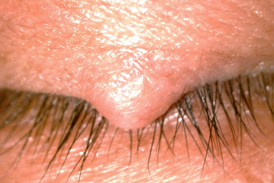
Figure 1. Clinical photograph of the right eye. Preoperative photograph shows a 4 × 3 mm-sized non-pigmented chala-zion-like mass on the right upper eyelid margin.
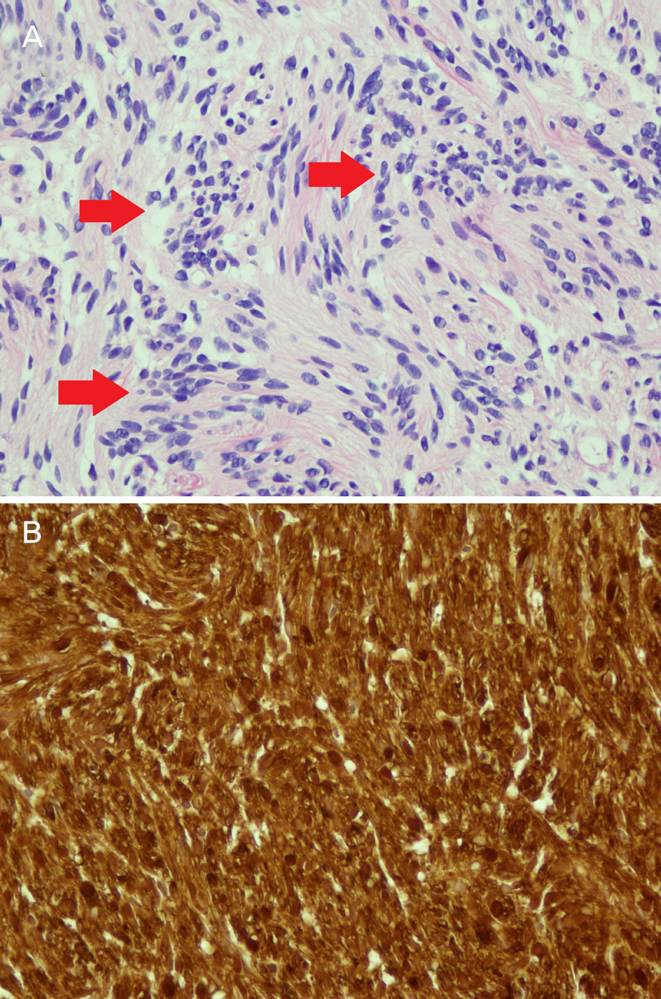
Figure 2. Light micrograph of excisional biopsy specimen. (A) The Antoni type A area shows cellular proliferation of spindle cells arranged in a palisading fashion (red arrows). In this case, type B area is not identified (hematoxylin-eosin, ×400). (B) Immunostaining for S100 protein is strong and dif-fuse (S-100, ×400).
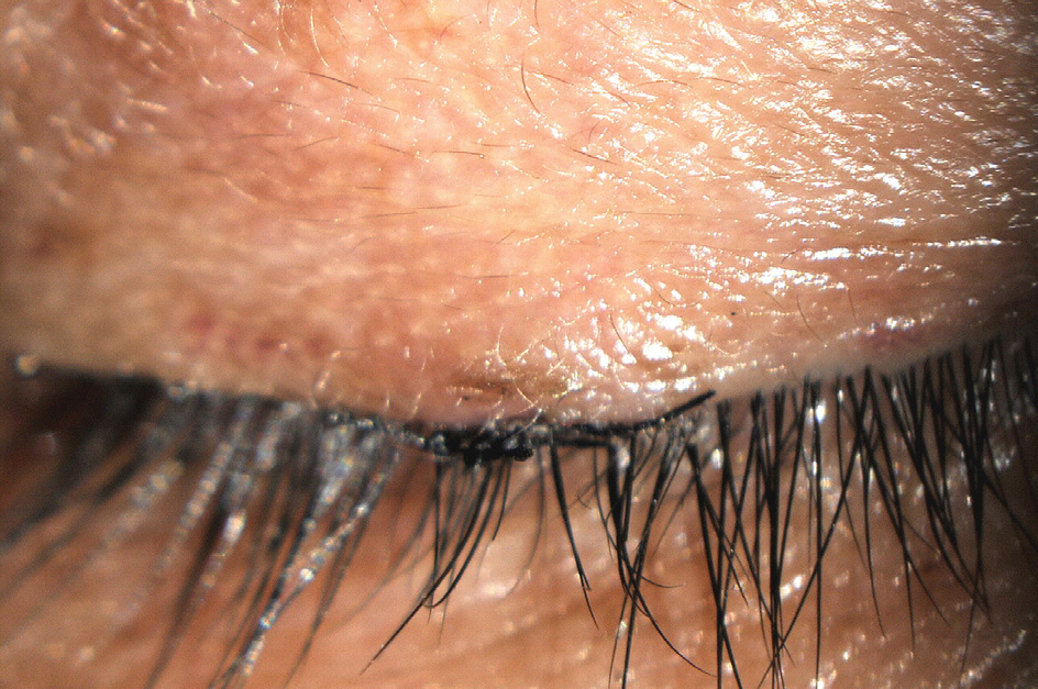
Figure 3. Clinical photograph of the right eye at 8 weeks after excision. Postoperative photograph showed removed site was clear. No recurrence was observed.
Reference
-
References
1. Welch, RB, Duke JR. Lesions of the lids: a statistical note. Am J Ophthalmol. 1961; 66:111–24.2. Allington HV, Allington JH. Eyelid tumors. Arch Dermatol. 1961; 66:111–24.
Article3. Sidiqui MA, Leslie T, Scott C, Mackenzie J. Eyelid schwannoma in a male adult. Clin Exp Ophthalmol. 1961; 66:111–24.
Article4. Chung YR, Moon S, Jang JW. Eyelid schwannoma in a Korean woman. Jpn J Ophthalmol. 1961; 66:111–24.
Article5. Baijal GC, Garg SK, Kanhere S, Monga S. Schwannoma of the eye-lid. Indian J Ophthalmol. 1961; 66:111–24.6. Butt Z, Ironside JW. Superficial epitheloid schwannoma present-ing as a subcutaneous upper eyelid mass. Br J Ophthalmol. 1961; 66:111–24.7. Gelincik I. Right upper eyelid schwannoma in a child: a case report. J Clin Exp Pathol. 2012; 2:1000116.
Article8. Shields JA, Guibor P. Neurilemoma of the eyelid resembling a re-current chalazion. Arch Ophthalmol. 1984; 102:1650.
Article9. Shields JA, Kiratli H, Shields CL, et al. Schwannoma of the eyelid in a child. J Pediatr Ophthalmol Strabismus. 1961; 66:111–24.
Article10. López-Tizón E, Mencía-Gutiérrez E, Gutiérrez-Díaz E, Ricoy JR. Schwannoma of the eyelid: report of two cases. Dermatol Online J. 2007; 13:12.
Article11. Lee KW, Lee MJ, Kim NJ, et al. A case of eyelid schwannoma. J Korean Ophthalmol Soc. 1961; 66:111–24.
Article12. Yuichi O, Tatsuo Y. A case of schwannoma in the upper eyelid. J Eye Ophthalmic Plast Reconstr Surg. 1961; 66:111–24.13. Kumar S, Kumar S, Kulshrestha R. Cystic schwannoma of eyelid in an Indian male: a rare presentation. Orbit. 1961; 66:111–24.
Article14. Singh S, Saraf S, Goswami D, Singh S. Case report of isolated schwannoma‑ a rare eyelid tumor. US Ophthalmic Review,. 2014; 7:143–5.15. Magdum RM, Paranjpe R, Kotecha M, Patil P. Solitary eyelid schwannoma. Med J DY Patil Univ. 1961; 66:111–24.
Article16. Rootman J, Goldberg C, Robertson W. Primary orbital schwan-nomas. Br J Ophthalmol. 1961; 66:111–24.
Article17. Le Marc’hadour F, Romanet JP, Fdili A, et al. Schwannoma of the bulbar conjunctiva. Arch Ophthalmol. 1961; 66:111–24.
Article18. Shields JA, Font RL, Eagle RC Jr. . Melanotic schwannoma of the choroid: immunohistochemistry and electron microscopic observations. Ophthalmology. 1961; 66:111–24.19. Graham CM, McCartney AC, Buckley RJ. Intrascleral neuril-emmoma. Br J Ophthalmol. 1961; 66:111–24.
Article
