J Korean Assoc Oral Maxillofac Surg.
2016 Dec;42(6):383-387. 10.5125/jkaoms.2016.42.6.383.
Verruciform xanthoma in the hard palate: a case report and literature review
- Affiliations
-
- 1Department of Surgery, Stomatology, Pathology and Radiology, Bauru School of Dentistry, University of São Paulo, Bauru, Brazil. alexsg@fob.usp.br
- KMID: 2364008
- DOI: http://doi.org/10.5125/jkaoms.2016.42.6.383
Abstract
- Oral verruciform xanthoma (OVX) is an uncommon lesion that appears on the oral mucosa. The aim of this paper was to discuss the probable etiopathogenesis of OVX in the hard palate, reinforcing the importance of including this lesion in the differential diagnosis of verrucous lesions. A 43-year-old male smoker presented with a painless lesion with a verrucous surface and erythematous spots on the hard palate. Excisional biopsy revealed oral mucosa consisting of hyperkeratosis, acanthosis, and elongated rete pegs. Subjacent connective tissue showed numerous foam cells with clear cytoplasm and pyknotic nucleus, negative on periodic acid-Schiff staining. Immunohistochemical analysis revealed foam cells positive for anti-CD68 antibody, while anti-KI-67 antibody was restricted to the basal layer of the oral epithelium. A final diagnosis of OVX was established. The patient showed no signs of recurrence after seven months of follow-up. Physical trauma and smoking habits can be directly related to the etiology of verruciform xanthoma because the lesion is chronic and inflammatory with slow growth, and sites if high trauma are more often affected by such a lesion. The hard palate is the second most commonly affected site, and local trauma caused by smoking can be a cause of this type of lesion.
Keyword
MeSH Terms
Figure
-
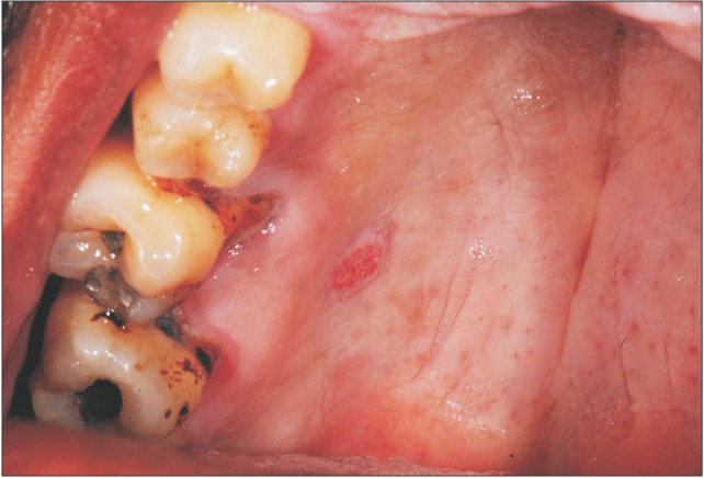
Fig. 1 Clinical appearance of the lesion on the hard palate showing verrucous surface and erythematous spots, measuring approximately 5 mm in diameter.
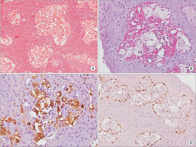
Fig. 2 Histopathological features of the verruciform xanthoma showing hyperkeratosis, acanthosis, elongated rete pegs and numerous foam cells with clear cytoplasm and pyknotic nucleus in the connective tissue (H&E staining, ×200; A), foam cells showing negative for periodic acid-Schiff (PAS staining, ×400; B), foam cells positive for anti-CD68 antibody (anti-CD68 staining, ×400; C), basal layer of the oral epithelium positive to KI-67 and negative for foam cells (anti-KI-67 staining, ×200; D).
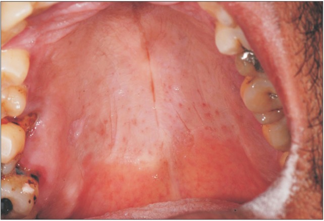
Fig. 3 Seven months of follow-up, no recurrences.
Reference
-
1. Shafer WG. Verruciform xanthoma. Oral Surg Oral Med Oral Pathol. 1971; 31:784–789. PMID: 5280461.
Article2. Hegde U, Doddawad VG, Sreeshyla H, Patil R. Verruciform xanthoma: a view on the concepts of its etiopathogenesis. J Oral Maxillofac Pathol. 2013; 17:392–396. PMID: 24574658.
Article3. Zegarelli DJ, Zegarelli-Schmidt EC, Zegarelli EV. Verruciform xanthoma. Further light and electron microscopic studies, with the addition of a third case. Oral Surg Oral Med Oral Pathol. 1975; 40:246–256. PMID: 1057149.4. Philipsen HP, Reichart PA, Takata T, Ogawa I. Verruciform xanthoma--biological profile of 282 oral lesions based on a literature survey with nine new cases from Japan. Oral Oncol. 2003; 39:325–336. PMID: 12676251.
Article5. Iamaroon A, Vickers RA. Characterization of verruciform xanthoma by in situ hybridization and immunohistochemistry. J Oral Pathol Med. 1996; 25:395–400. PMID: 8890055.
Article6. Nowparast B, Howell FV, Rick GM. Verruciform xanthoma. A clinicopathologic review and report of fifty-four cases. Oral Surg Oral Med Oral Pathol. 1981; 51:619–625. PMID: 6942361.7. Neville B. The verruciform xanthoma. A review and report of eight new cases. Am J Dermatopathol. 1986; 8:247–253. PMID: 3728885.
Article8. Kakarantza-Angelopoulou E, Nicolatou O, Anagnostopoulou S. Verruciform xanthoma of the palate: case report with electron microscopy. J Oral Maxillofac Surg. 1991; 49:409–412. PMID: 2005497.9. Mostafa KA, Takata T, Ogawa I, Ijuhin N, Nikai H. Verruciform xanthoma of the oral mucosa: a clinicopathological study with immunohistochemical findings relating to pathogenesis. Virchows Arch A Pathol Anat Histopathol. 1993; 423:243–248. PMID: 8236821.
Article10. Rawal SY, Kalmar JR, Tatakis DN. Verruciform xanthoma: immunohistochemical characterization of xanthoma cell phenotypes. J Periodontol. 2007; 78:504–509. PMID: 17335374.
Article11. Buchner A, Hansen LS, Merrell PW. Verruciform xanthoma of the oral mucosa. Report of five cases and review of the literature. Arch Dermatol. 1981; 117:563–565. PMID: 7294847.
Article12. Hume WJ, Smith CJ, Franklin CD. Verruciform xanthoma. Br J Oral Surg. 1980; 18:157–161. PMID: 6934808.
Article13. Neville BW, Weathers DR. Verruciform xanthoma. Oral Surg Oral Med Oral Pathol. 1980; 49:429–434. PMID: 6154914.
Article14. Rhinow K, Kalz S, Gelderblom R, Reichart A. Verruciform xanthoma. Mund Kiefer Gesichtschir. 2003; 7:52–55. PMID: 12556987.15. Hu JA, Li Y, Li S. Verruciform xanthoma of the oral cavity: clinicopathological study relating to pathogenesis. Report of three cases APMIS. 2005; 113:629–634. PMID: 16218939.16. Yu CH, Tsai TC, Wang JT, Liu BY, Wang YP, Sun A, et al. Oral verruciform xanthoma: a clinicopathologic study of 15 cases. J Formos Med Assoc. 2007; 106:141–147. PMID: 17339158.
Article17. Cheng YS, Wright J, Lucente J, McQuade MJ. Oral and maxillofacial pathology case of the month. Verruciform xanthoma. Tex Dent J. 2010; 127:126–127. 130–131. PMID: 20162945.18. Shahrabi Farahani S, Treister NS, Khan Z, Woo SB. Oral verruciform xanthoma associated with chronic graft-versus-host disease: a report of five cases and a review of the literature. Head Neck Pathol. 2011; 5:193–198. PMID: 21305367.19. Ryu DJ, Lee SH, Yuk JI, Kim HJ, Huh JK, Park KH. Verruciform xanthoma of the palatal gingiva: a report of two cases. J Korean Assoc Oral Maxillofac Surg. 2013; 39:292–296. PMID: 24516820.
Article20. Aggarwal S, Aggarwal A, Gill S, Bakshi Y, Singh HP. Verruciform xanthoma of oral cavity- a case report. J Clin Diagn Res. 2014; 8:FD11–FD12.21. Bhattacharyya I, Islam N. Diagnostic discussion. Verruciform xanthoma. Todays FDA. 2014; 26:49–51. PMID: 24988733.22. Tang R, Kopp SA, Cobb C, Halpern AV. Disseminated verruciform xanthoma: a case report. Cutis. 2014; 93:307–310. PMID: 24999643.23. Ide F, Obara K, Yamada H, Mishima K, Saito I, Kusama K. Cellular basis of verruciform xanthoma: immunohistochemical and ultrastructural characterization. Oral Dis. 2008; 14:150–157. PMID: 18302675.
Article24. Sah K, Kale AD, Hallikerimath S. Verruciform xanthoma: report of two cases and review on pathogenesis. J Oral Maxillofac Pathol. 2008; 12:41–44.
Article
- Full Text Links
-
- Actions
-
Cited
- CITED
-
- Close
- Share
-
- Similar articles
-
- A Case of Verruciform Genital-associated (Vegas) Xanthoma on the Scrotum
- Clinical, histological, and deep learning-based assessments and treatment of oral verruciform xanthoma: a case report
- Verruciform Xanthoma of the Penis
- A Case of Verruciform Xanthoma Associated with Epidermal Atypia
- A Case of Verruciform Xanthoma Presenting as Leukoplakia on the Lower Lip
