Prospective randomized clinical trial of hydrophilic tapered implant placement at maxillary posterior area: 6 weeks and 12 weeks loading
- Affiliations
-
- 1Department of Oral and Maxillofacial Surgery, Section of Dentistry, Seoul National University Bundang Hospital, Seongnam, Republic of Korea. kyk0505@snubh.org
- 2Department of Prosthodontics, Section of Dentistry, Seoul National University Bundang Hospital, Seongnam, Republic of Korea.
- 3Department of Dentistry & Dental Research Institute, School of Dentistry, Seoul National University, Seoul, Republic of Korea.
- 4Department of Science Education, College of Education, Dankook University, Yongin, Republic of Korea.
- KMID: 2355444
- DOI: http://doi.org/10.4047/jap.2016.8.5.396
Abstract
- PURPOSE
Early loading of implant can be determined by excellent primary stability and characteristic of implant surface. The implant system with recently improved surface can have load application 4-6 weeks after installing in maxilla and mandible. This study evaluated the effect of healing period to the stability of hydrophilic tapered-type implant at maxillary posterior area.
MATERIALS AND METHODS
This study included 30 patients treated by hydrophilic tapered-type implants (total 41 implants at maxilla) and classified by two groups depending on healing period. Group 1 (11 patients, 15 implants) was a control group and the healing period was 12 weeks, and Group 2 (19 patients, 26 implants) was test group and the healing period was 6 weeks. Immediately after implant placement, at the first impression taking, implant stability was measured using Osstell Mentor. The patients also took periapical radiographs after restoration delivery, 12 months after restoration and final followup period. The marginal bone loss around the implants was measured using the periapical radiographs.
RESULTS
All implants were survived and success rate was 97.56%. The marginal bone loss was less than 1mm after 1 year postoperatively except the one implant. The stabilities of the implants were not correlated with age, healing period until loading, insertion torque (IT), the diameter of fixture and the location of implant. Only the quality of bone in group 2 (6 week) was correlated with the stability of implant.
CONCLUSION
Healing period of 6 weeks can make the similar clinical prognosis of implants to that of healing period of 12 weeks if bone quality is carefully considered in case of early loading.
Keyword
MeSH Terms
Figure
-
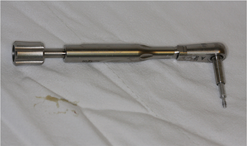
Fig. 1 Osstell SmartPeg was connected with 5 Ncm using hand torque wrench.
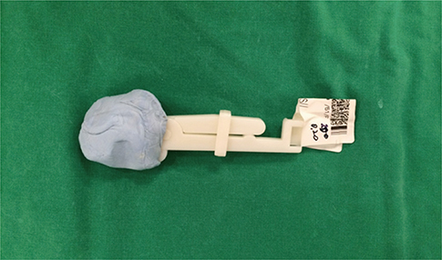
Fig. 2 Film positioner with putty jig.
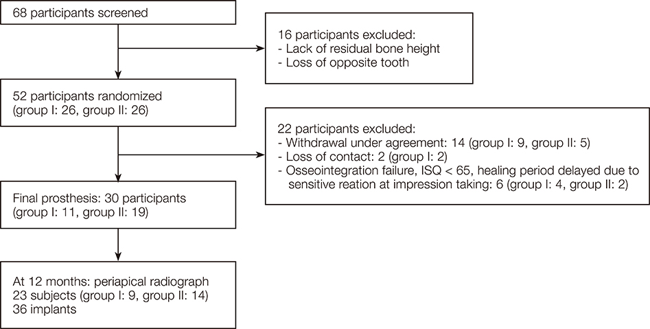
Fig. 3 CONSORT study flow chart.
Cited by 2 articles
-
Comparison of sandblasted and acid-etched surface implants and new hydrophilic surface implants in the posterior maxilla using a 3-month early-loading protocol: a randomized controlled trial
Hyeong Gi Kim, Pil-Young Yun, Young-Kyun Kim, Il-hyung Kim
J Korean Assoc Oral Maxillofac Surg. 2021;47(3):175-182. doi: 10.5125/jkaoms.2021.47.3.175.Randomized clinical trial to evaluate the efficacy and safety of two types of sandblasted with large-grit and acid-etched surface implants with different surface roughness
Jun-Hyung Jeon, Min-Joong Kim, Pil-Young Yun, Deuk-Won Jo, Young-Kyun Kim
J Korean Assoc Oral Maxillofac Surg. 2022;48(4):225-231. doi: 10.5125/jkaoms.2022.48.4.225.
Reference
-
1. Albrektsson T, Brånemark PI, Hansson HA, Lindström J. Osseointegrated titanium implants. Requirements for ensuring a long-lasting, direct bone-to-implant anchorage in man. Acta Orthop Scand. 1981; 52:155–170.2. Babbush CA. Titanium plasma spray screw implant system for reconstruction of the edentulous mandible. Dent Clin North Am. 1986; 30:117–131.3. Brånemark PI, Hansson BO, Adell R, Breine U, Lindström J, Hallén O, Ohman A. Osseointegrated implants in the treatment of the edentulous jaw. Experience from a 10-year period. Scand J Plast Reconstr Surg Suppl. 1977; 16:1–132.4. Cochran DL, Buser D, ten Bruggenkate CM, Weingart D, Taylor TM, Bernard JP, Peters F, Simpson JP. The use of reduced healing times on ITI implants with a sandblasted and acid-etched (SLA) surface: early results from clinical trials on ITI SLA implants. Clin Oral Implants Res. 2002; 13:144–153.5. Abrahamsson I, Berglundh T, Linder E, Lang NP, Lindhe J. Early bone formation adjacent to rough and turned endosseous implant surfaces. An experimental study in the dog. Clin Oral Implants Res. 2004; 15:381–392.6. Choi JM, Kim HE, Lee IS. Ion-beam-assisted deposition (IBAD) of hydroxyapatite coating layer on Ti-based metal substrate. Biomaterials. 2000; 21:469–473.7. Kim H, Choi SH, Chung SM, Li LH, Lee IS. Enhanced bone forming ability of SLA-treated Ti coated with a calcium phosphate thin film formed by e-beam evaporation. Biomed Mater. 2010; 5:044106.8. Kim H, Choi SH, Chung SM, Li LH, Lee IS. Enhanced bone forming ability of SLA-treated Ti coated with a calcium phosphate thin film formed by e-beam evaporation. Biomed Mater. 2010; 5:044106.9. Ganeles J, Zöllner A, Jackowski J, ten Bruggenkate C, Beagle J, Guerra F. Immediate and early loading of Straumann implants with a chemically modified surface (SLActive) in the posterior mandible and maxilla: 1-year results from a prospective multicenter study. Clin Oral Implants Res. 2008; 19:1119–1128.10. Zöllner A, Ganeles J, Korostoff J, Guerra F, Krafft T, Brägger U. Immediate and early non-occlusal loading of Straumann implants with a chemically modified surface (SLActive) in the posterior mandible and maxilla: interim results from a prospective multicenter randomized-controlled study. Clin Oral Implants Res. 2008; 19:442–450.11. Bornstein MM, Schmid B, Belser UC, Lussi A, Buser D. Early loading of non-submerged titanium implants with a sand-blasted and acid-etched surface. 5-year results of a prospective study in partially edentulous patients. Clin Oral Implants Res. 2005; 16:631–638.12. Friberg B, Sennerby L, Linden B, Gröndahl K, Lekholm U. Stability measurements of one-stage Brånemark implants during healing in mandibles. A clinical resonance frequency analysis study. Int J Oral Maxillofac Surg. 1999; 28:266–272.13. Noguerol B, Muñoz R, Mesa F, de Dios Luna J, O'Valle F. Early implant failure. Prognostic capacity of Periotest: retrospective study of a large sample. Clin Oral Implants Res. 2006; 17:459–464.14. Atieh MA, Alsabeeha NH, Payne AG. Can resonance frequency analysis predict failure risk of immediately loaded implants? Int J Prosthodont. 2012; 25:326–339.15. Ribeiro-Rotta RF, de Oliveira RC, Dias DR, Lindh C, Leles CR. Bone tissue microarchitectural characteristics at dental implant sites part 2: correlation with bone classification and primary stability. Clin Oral Implants Res. 2014; 25:e47–e53.16. Park KJ, Kwon JY, Kim SK, Heo SJ, Koak JY, Lee JH, Lee SJ, Kim TH, Kim MJ. The relationship between implant stability quotient values and implant insertion variables: a clinical study. J Oral Rehabil. 2012; 39:151–159.17. Makary C, Rebaudi A, Sammartino G, Naaman N. Implant primary stability determined by resonance frequency analysis: correlation with insertion torque, histologic bone volume, and torsional stability at 6 weeks. Implant Dent. 2012; 21:474–480.18. Lekholm U, Zarb GA. Patient selection and preparation. In : Brånemark PI, Zarb GA, Albrektsson T, editors. Tissue Integrated Prostheses: Osseointegration in Clinical Dentistry. Chicago: Quintessence Publ Co;1985. p. 199–209.19. Zarb GA, Albrektsson T. Consensus report: towards optimized treatment outcomes for dental implants. J Prosthet Dent. 1998; 80:641.20. Sennerby L, Meredith N. Analisi della freuqenza di resonanza(RFA). Conoscenze attuali e implicazioni cliniche. In : Chipasco M, Gatti C, editors. Osteointegrazione e Carico Immediato. Fondamenti Biologici e Applicazioni Cliniche. Milan: Masson;2002. p. 19–31.21. Kessler-Liechti G, Zix J, Mericske-Stern R. Stability measurements of 1-stage implants in the edentulous mandible by means of resonance frequency analysis. Int J Oral Maxillofac Implants. 2008; 23:353–358.22. Ostman PO, Hellman M, Wendelhag I, Sennerby L. Resonance frequency analysis measurements of implants at placement surgery. Int J Prosthodont. 2006; 19:77–83.23. Bilhan H, Geckili O, Mumcu E, Bozdag E, Sünbüloğlu E, Kutay O. Influence of surgical technique, implant shape and diameter on the primary stability in cancellous bone. J Oral Rehabil. 2010; 37:900–907.24. Park JH, Lim YJ, Kim MJ, Kwon HB. The effect of various thread designs on the initial stability of taper implants. J Adv Prosthodont. 2009; 1:19–25.25. Ahn SJ, Leesungbok R, Lee SW, Heo YK, Kang KL. Differences in implant stability associated with various methods of preparation of the implant bed: an in vitro study. J Prosthet Dent. 2012; 107:366–372.26. Bayarchimeg D, Namgoong H, Kim BK, Kim MD, Kim S, Kim TI, Seol YJ, Lee YM, Ku Y, Rhyu IC, Lee EH, Koo KT. Evaluation of the correlation between insertion torque and primary stability of dental implants using a block bone test. J Periodontal Implant Sci. 2013; 43:30–36.27. Degidi M, Daprile G, Piattelli A. Determination of primary stability: a comparison of the surgeon's perception and objective measurements. Int J Oral Maxillofac Implants. 2010; 25:558–561.28. Pommer B, Hof M, Fädler A, Gahleitner A, Watzek G, Watzak G. Primary implant stability in the atrophic sinus floor of human cadaver maxillae: impact of residual ridge height, bone density, and implant diameter. Clin Oral Implants Res. 2014; 25:e109–e113.29. Ribeiro-Rotta RF, de Oliveira RC, Dias DR, Lindh C, Leles CR. Bone tissue microarchitectural characteristics at dental implant sites part 2: correlation with bone classification and primary stability. Clin Oral Implants Res. 2014; 25:e47–e53.30. Marquezan M, Osório A, Sant'Anna E, Souza MM, Maia L. Does bone mineral density influence the primary stability of dental implants? A systematic review. Clin Oral Implants Res. 2012; 23:767–774.31. Isoda K, Ayukawa Y, Tsukiyama Y, Sogo M, Matsushita Y, Koyano K. Relationship between the bone density estimated by cone-beam computed tomography and the primary stability of dental implants. Clin Oral Implants Res. 2012; 23:832–836.32. Farré-Pagés N, Augé-Castro ML, Alaejos-Algarra F, Mareque-Bueno J, Ferrés-Padró E, Hernández-Alfaro F. Relation between bone density and primary implant stability. Med Oral Patol Oral Cir Bucal. 2011; 16:e62–e67.33. Hong J, Lim YJ, Park SO. Quantitative biomechanical analysis of the influence of the cortical bone and implant length on primary stability. Clin Oral Implants Res. 2012; 23:1193–1197.34. Hsu JT, Fuh LJ, Tu MG, Li YF, Chen KT, Huang HL. The effects of cortical bone thickness and trabecular bone strength on noninvasive measures of the implant primary stability using synthetic bone models. Clin Implant Dent Relat Res. 2013; 15:251–261.
- Full Text Links
-
- Actions
-
Cited
- CITED
-
- Close
- Share
-
- Similar articles
-
- One-staged sinus bone graft using tapered porous surfaced implant in lower residual bone height of maxillary sinus
- Fixation of the Sacroiliac Joint: A Cadaver-Based Concurrent-Controlled Biomechanical Comparison of Posterior Interposition and Posterolateral Transosseous Techniques
- The effect of loading time on the stability of mini-implant
- Guidance and rationale for the immediate implant placement in the maxillary molar
- Retrospective clinical study on sinus bone graft and tapered-body implant placement
