J Korean Ophthalmol Soc.
2014 Jan;55(1):129-132.
Delayed-Onset Interface Fluid Syndrome after LASIK Surgery in Traumatic Hyphema
- Affiliations
-
- 1HanGil Eye Hospital, Incheon, Korea.
- 2Department of Ophthalmology, Asan Medical Center, University of Ulsan College of Medicine, Seoul, Korea.
- 3Kimkisu Eye Clinic, Jeju, Korea. oijee@hanmail.net
Abstract
- A 50-year-old female was referred to our clinic with visual disturbance, hyphema and increased intraocular pressure (IOP) in her right eye 7 days after experiencing blunt trauma in that eye. She had undergone uncomplicated laser in situ keratomileusis (LASIK) on both eyes 10 years earlier. At initial examination, the best corrected visual acuity (BCVA) in her right eye was counting fingers at 2 feet with no correction. Central Goldmann applanation tonometry (GAT) showed an IOP of 7 mm Hg. Peripheral digital tonometry showed the IOPs in her right eye superiorly, nasally, temporally, and inferiorly were 36 mm Hg, 35 mm Hg, 34.5 mm Hg and 36.5 mm Hg, respectively. Slit-lamp examination showed diffuse epithelial and stromal edema and a blood clot 1 mm in height in the anterior chamber. Spectral domain scanning laser ophthalmoscope/optical coherence tomography (SD-SLO/OCT) images showed a pocket of fluid between the LASIK flap and the underlying stroma. The patient was started on anti-inflammatory agent and IOP lowering agents. After 15 days of treatment, IOP measured with GAT was 10 mm Hg, slit-lamp examination showed that epithelial and stromal edema had disappeared, and OCT showed no fluid between the corneal flap and stroma.
MeSH Terms
Figure
-
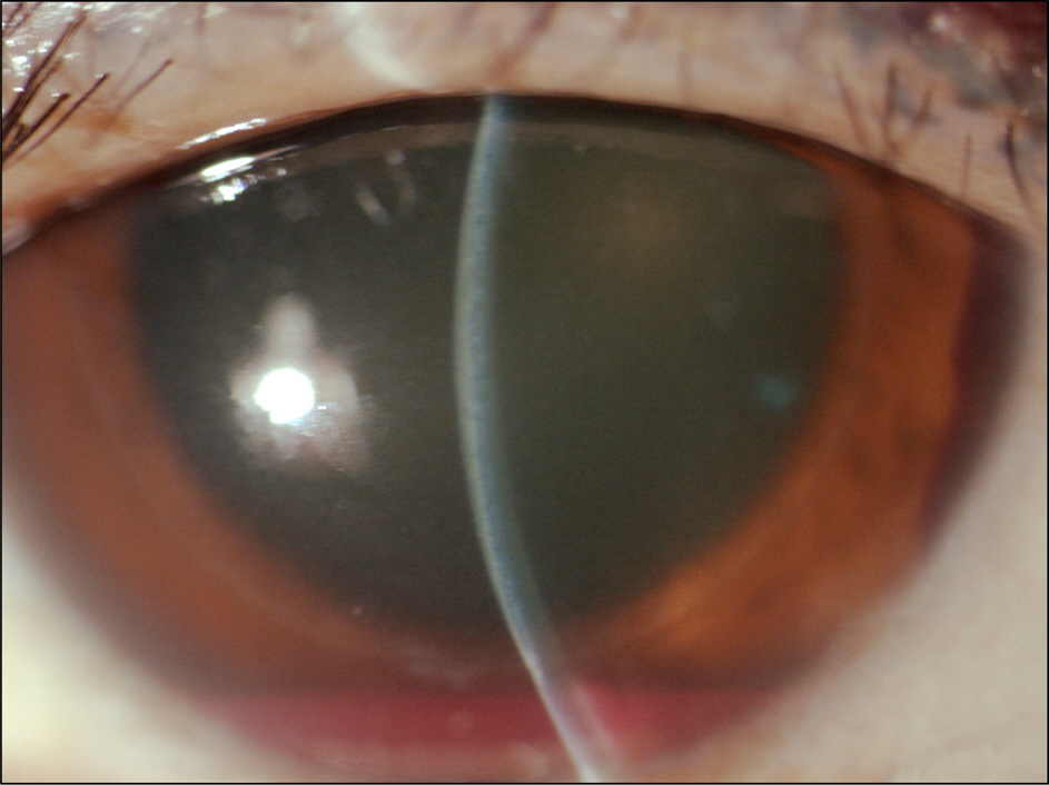
Figure 1. Slit-lamp examination showing diffuse epithelial and stromal edema, and 1 mm height blood clot 1 mm height in the anterior chamber of the right eye.
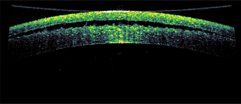
Figure 2. SD-SLO/OCT images of the cornea of the right eye with central edema and a fluid-filled pocket at the LASIK interface.
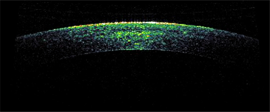
Figure 3. OCT images of cornea after 9 days of treatment, showing a decrease in interface fluid.
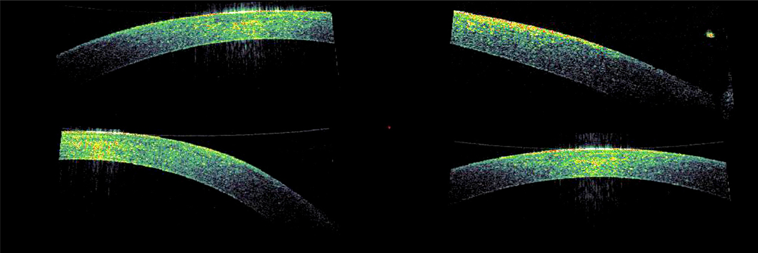
Figure 4. OCT images after 15 days of treatment, interface fluid is not observed.
Reference
-
References
1. Sugar A, Rapuano CJ, Culbertson WW. . Laser in situ kerato-mileusis for myopia and astigmatism: safety and efficacy: a report by the American Academy of Ophthalmology. Ophthalmology. 2002; 109:175–87.2. Schallhorn SC, Amesbury EC, Tanzer DJ.Avoidance, recognition, and management of LASIK complications. Am J Ophthalmol. 2006; 141:733–9.
Article3. Lin RT, Maloney RK.Flap complications associated with lamellar refractive surgery. Am J Ophthalmol. 1999; 127:129–36.
Article4. Lyle WA, Jin GJ.Interface fluid associated with diffuse lamellar keratitis and epithelial ingrowth after laser in situ keratomileusis. J Cataract Refract Surg. 1999; 25:1009–12.
Article5. Hamilton DR, Manche EE, Rich LF, Maloney RK.Steroid-induced glaucoma after laser in situ keratomileusis associated with inter-face fluid. Ophthalmology. 2002; 109:659–65.
Article6. Hoffman RS, Fine IH, Packer M.Persistent interface fluid syndrome. J Cataract Refract Surg. 2008; 34:1405–8.
Article7. McLeod SD, Mather R, Hwang DG, Margolis TP.Uveitis-associated flap edema and lamellar interface fluid collection after LASIK. Am J Ophthalmol. 2005; 139:1137–9.
Article8. Dawson DG, Schmack I, Holley GP. . Interface fluid syndrome in human eye bank corneas after LASIK: causes and pathogenesis. Ophthalmology. 2007; 114:1848–59.9. Kim JC, Chung TY, Kee CW.A wide discrepancy between intra-ocular pressure by applanation and non-contact tonometry after LASIK. J Korean Ophthalmol Soc. 2007; 48:860–5.10. Park CK, Kim JH.Comparison of wound healing after photo-refractive keratectomy and laser in situ keratomileusis in rabbits. J Cataract Refract Surg. 1999; 25:842–50.
Article11. Rehany U, Bersudsky V, Rumelt S.Paradoxical hypotony after laser in situ keratomileusis. J Cataract Refract Surg. 2000; 26:1823–6.
Article12. Han SB, Woo SJ, Hyon JY.Delayedonset interface fluid syndrome after laser in situ keratomileusis secondary to combined cataract and vitreoretinal surgery. J Cataract Refract Surg. 2012; 38:548–50.
Article13. Ortega-Usobiaga J, Martin-Reyes C, Llovet-Osuna F. . Interface fluid syndrome in routine cataract surgery 10 years after laser in situ keratomileusis. Cornea. 2012; 31:706–7.
Article
- Full Text Links
-
- Actions
-
Cited
- CITED
-
- Close
- Share
-
- Similar articles
-
- A Wide Discrepancy between Intraocular Pressure by Applanation and Non-contact Tonometry after LASIK
- LASIK Interface-Captured Foreign Bodies after Mild Traumatic Corneal Scratch without Flap Displacement
- A Clinical Study on the Traumatic Hyphema
- High Intraocular Pressure-induced Delayed Diffuse Lamellar Keratitis after Laser In Situ Keratomileusis (LASIK)
- A Clinical Study on Trauamtic Hyphema in Chunbuk area
