LASIK Interface-Captured Foreign Bodies after Mild Traumatic Corneal Scratch without Flap Displacement
- Affiliations
-
- 1Department of Ophthalmology and Visual Science, St. Vincent Hospital, The Catholic University of Korea College of Medicine, Seoul, Korea.
- 2Department of Ophthalmology and Visual Science, Seoul St. Mary's Hospital, The Catholic University of Korea College of Medicine, Seoul, Korea. mskim@catholic.ac.kr
- KMID: 1376130
- DOI: http://doi.org/10.3341/kjo.2012.26.3.222
Abstract
- A 38-year-old woman developed diffusely distributed opacities with crystalline materials in the laser in situ keratomileusis (LASIK) interface of her eye after she was scratched by a sprig during mountain climbing. No sign of flap displacement was noted. Despite two days of topical and systemic antibiotics therapy, the corneal infiltration with interface opacities persisted. The following day, the distribution of the crystalline materials had rotated in a counterclockwise direction. Flap lifting and foreign body removal using sufficient irrigation were performed. One month after surgery, the patient's postoperative uncorrected visual acuity was 0.8 with cleared interface. No signs of epithelial ingrowth or flap striae were noted. Mild traumatic corneal scratching without flap displacement may threaten the integrity of the LASIK interface. If foreign bodies are suspected to be the cause of inflammation, early flap lifting with irrigation is imperative for successful treatment.
MeSH Terms
Figure
-
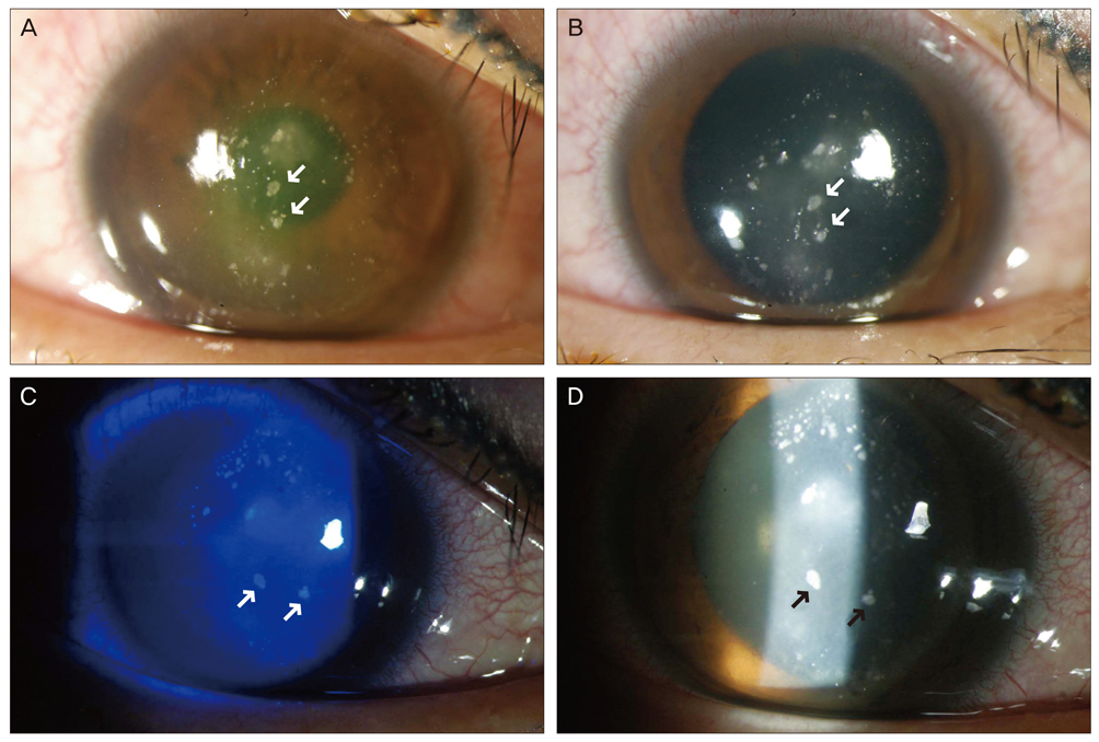
Fig. 1 Slit-lamp photograph of the patient before flap lifting and irrigation. Upper photographs (A) (before pupillary dilatation) and (B) (after papillary dilatation) show diffusely distributed crystalline materials with stromal infiltration at the laser in situ keratomileusis interface on the first day of hospitalization. Note three large materials distributed linearly in the center of the cornea (see arrows). On the third day of hospitalization, photographs (C) (under cobalt blue filter) and (D) (under slit beam) show the change of distribution of the crystalline materials. Note three large materials in the center that seem to have rotated counterclockwise.
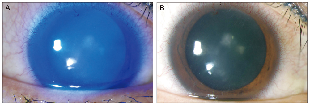
Fig. 2 Slit-lamp photograph of the patient after flap lifting and irrigation. In (A) and (B), crystalline materials at the laser in situ keratomileusis interface were cleared and conjunctival injection decreased on the first postoperative day.
Reference
-
1. Aldave AJ, Hollander DA, Abbott RL. Late-onset traumatic flap dislocation and diffuse lamellar inflammation after laser in situ keratomileusis. Cornea. 2002. 21:604–607.2. Lemley HL, Chodosh J, Wolf TC, et al. Partial dislocation of laser in situ keratomileusis flap by air bag injury. J Refract Surg. 2000. 16:373–374.3. Jin GJ, Merkley KH. Laceration and partial dislocation of LASIK flaps 7 and 4 years postoperatively with 20 / 20 visual acuity after repair. J Refract Surg. 2006. 22:904–905.4. Melki SA, Talamo JH, Demetriades AM, et al. Late traumatic dislocation of laser in situ keratomileusis corneal flaps. Ophthalmology. 2000. 107:2136–2139.5. Cheung LM, Papalkar D, Versace P. Traumatic late flap dehiscence and Enterobacter keratitis following LASIK. J Refract Surg. 2006. 22:402–404.6. Chang MA, Jain S, Azar DT. Infections following laser in situ keratomileusis: an integration of the published literature. Surv Ophthalmol. 2004. 49:269–280.7. Maurice DM, Monroe F. Cohesive strength of corneal lamellae. Exp Eye Res. 1990. 50:59–63.
- Full Text Links
-
- Actions
-
Cited
- CITED
-
- Close
- Share
-
- Similar articles
-
- Clinical feature of unintended thin corneal flap in LASIK: 1-year follow-up
- A Case of Acute Central Stromal Melting after LASIK
- LASIK Using the Manual Microkeratome: Complications, Management, and Result
- The Consistency of Corneal Flap Thickness and Size in LASIK using the Innovatome Automatic Micro keratome
- A Case of Mycobacterium Fortuitum Keratitis at the Interface of the Cornea after LASIK
