J Korean Ophthalmol Soc.
2012 Feb;53(2):353-356.
Moyamoya Disease Initially Presenting Transient Visual Loss
- Affiliations
-
- 1Department of Ophthalmology, Sanggye Paik Hospital, Inje University College of Medicine, Seoul, Korea.
- 2Department of Ophthalmology, Seoul Paik Hospital, Inje University College of Medicine, Seoul, Korea.
- 3Department of Ophthalmology, Armed Forces Capital Hospital, Seongnam, Korea. brainh@hanmail.net
Abstract
- PURPOSE
To report a case of moyamoya disease initially presenting transient visual loss in a healthy young subject.
CASE SUMMARY
A 20-year-old male with no history of systemic disease or trauma visited our clinic due to sudden onset visual loss in the right eye. There were no accompanying symptoms, including headache, seizure, paresis, or paresthesia. Best corrected visual acuity at the first visit was hand movement in the right eye and 20/20 in the left eye. No abnormal finding was revealed in the anterior segment. On fundus examination, whitening at post pole was found in the right eye. In fluorescein angiography, a choroidal and retinal artery filling delay in the posterior pole was noted. The patient's visual acuity began to improve gradually and was recovered to 20/20 by the next day. Moyamoya disease was diagnosed based on magnetic resonance angiography of the brain and transfemoral cerebral angiography as well as stenosis of the internal carotid artery and middle cerebral artery with collateral vessel networks.
CONCLUSIONS
Moyamoya disease should be considered as a possible cause of transient visual loss in healthy young subjects.
Keyword
MeSH Terms
Figure
-
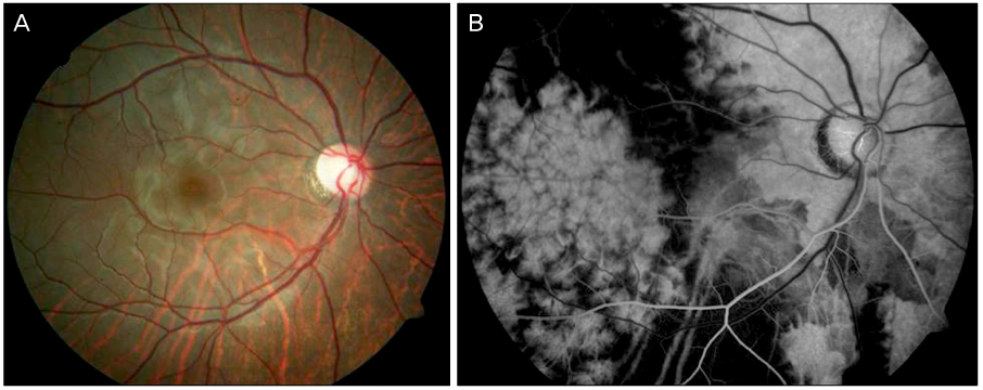
Figure 1 (A) In fundus photograph of the right eye at first visit, retinal opacification veiling choroidal vasculature was found. (B) In fluorescein angiography, filling defects and prominent watershed zone of choroid in posterior pole and delayed retinal arterial filling were revealed.
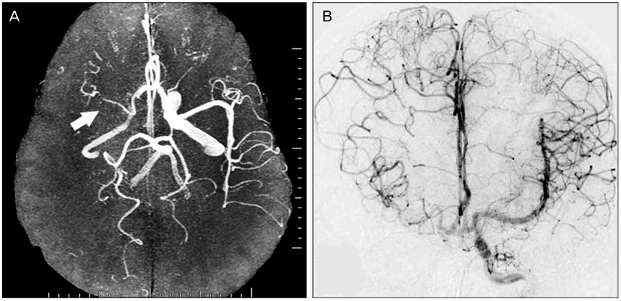
Figure 2 Magnetic resonance angiography of brain (A) and transfemoral cerebral angiography (B) showed typical findings of moyamyoa disease; stenosis of internal carotid artery and middle cerebral artery (white arrow of figure A) with collateral vessels (B).
Reference
-
1. Suzuki J, Kodama N. Moyamoya disease--a review. Stroke. 1983. 14:104–109.2. Slamovits TL, Klingele TG, Burde RM, Gado MH. Moyamoya disease with central retinal vein occlusion. Case report. J Clin Neuroophthalmol. 1981. 1:123–127.3. Chace R, Hedges TR 3rd. Retinal artery occlusion due to moyamoya disease. J Clin Neuroophthalmol. 1984. 4:31–34.4. Noda S, Hayasaka S, Setogawa T, Matsumoto S. Ocular symptoms of moyamoya disease. Am J Ophthalmol. 1987. 103:812–816.5. Ushimura S, Mochizuki K, Ohashi M, et al. Sudden blindness in the fourth month of pregnancy led to diagnosis of moyamoya disease. Ophthalmologica. 1993. 207:169–173.6. Barrall JL, Summers CG. Ocular ischemic syndrome in a child with moyamoya disease and neurofibromatosis. Surv Ophthalmol. 1996. 40:500–504.7. Massaro M, Thorarensen O, Liu GT, et al. Morning glory disc anomaly and moyamoya vessels. Arch Ophthalmol. 1998. 116:253–254.8. Kim YS, Lee KS, Kim SH, Byun YJ. Moyamoya disease with characteristic fundus findings of retinal vascular insufficiency. J Korean Ophthalmol Soc. 1998. 39:2477–2483.9. Kim SH, Kim SY, Hong SB, et al. Two cases of moyamoya disease showing visal disturbance and complete occlusion of proximal internal carotid artery. J Korean Neurol Assoc. 1990. 8:325–333.10. Kaneko A, Irino S, Tomioka R, et al. Lower altitudinal bilateral hemianopsia in a patient with moyamoya disease. J Neurol. 2000. 247:383–384.11. Chu MK, Lee IH, Kim DI, Kim SM. Moyamoya disease initially presenting visual field defect. Yonsei Med J. 2001. 42:566–570.12. Harissi-Dagher M, Sebag M, Dagher JH, Moumdjian R. Chorioretinal atrophy in a patient with moyamoya disease. Case report. J Neurosurg. 2004. 101:843–845.13. Kim JH, Kim JY. A case of ophthalmic artery occlusion in moyamoya disease. J Korean Ophthalmol Soc. 2007. 48:849–853.14. Chen CS, Lee AW, Kelman S, Wityk R. Anterior ischemic optic neuropathy in moyamoya disease: a first case report. Eur J Neurol. 2007. 14:823–825.15. Kim DS, Kang SG, Yoo DS, et al. Sudden cortical blindness in an adult with moyamoya disease. Surg Neurol. 2007. 67:303–307.16. Grosberg BM, Solomon S, Friedman DI, Lipton RB. Retinal migraine reappraised. Cephalalgia. 2006. 26:1275–1286.17. O'Sullivan F, Rossor M, Elston JS. Amaurosis fugax in young people. Br J Ophthalmol. 1992. 76:660–662.
- Full Text Links
-
- Actions
-
Cited
- CITED
-
- Close
- Share
-
- Similar articles
-
- Moyamoya Disease Initially Presenting Visual Field Defect
- Reversible Cerebral Vasoconstriction Syndrome Misdiagnosed as Moyamoya Disease with Transient Ischemic Attack as Initial Manifestation
- A Case of Moyamoya Disease without Transient Ischemic Attacks
- A Case of Moyamoya Disease Initially Presenting as Anterior Ischemic Optic Neuropathy
- Anesthesia for a Patient with Moyamoya Disease presenting for Emergency Cesarean Section: A case report
