J Periodontal Implant Sci.
2011 Apr;41(2):98-104.
Case series of maxillary sinus augmentation with biphasic calcium phosphate: a clinical and radiographic study
- Affiliations
-
- 1Department of Periodontology, Research Institute for Periodontal Regeneration, Yonsei University College of Dentistry, Seoul, Korea. shchoi726@yuhs.ac
Abstract
- PURPOSE
The aim of this study was to evaluate 3.5 years-cumulative survival rate of implants placed on augmented sinus using Osteon, a bone graft material, and to assess the height of the grafted material through radiographic evaluation.
METHODS
Twenty patients were treated with maxillary sinus augmentation and 45 implant fixtures were installed simultaneously or after 6 months healing period. The height of the augmented sinus and the loss of marginal bone were measured by panoramic and intraoral radiographs immediately after augmentation and up to 42 months (mean, 19.4 months) subsequently. Changes in the height of the sinus graft material were calculated radiographically.
RESULTS
The cumulative survival rate was 95.56% in all 45 implants. Additionally, normal healing process without any complication was observed in all patients. The original sinus height was mean 4.3 mm and the augmented sinus height was mean 13.4 mm after the surgery. The mean marginal bone loss till 42 months was 0.52+/-0.56 mm. The reduced height of Osteon was 0.83+/-0.38 mm and it did not show significant correlation with the follow up periods (P=0.102). There were no statistically significant differences in reduced height of Osteon according to the simultaneous/delayed implantation (P=0.299) and particle size of Osteon (P=0.644).
CONCLUSIONS
It can be suggested that Osteon may have predictable result when it was used as a grafting material for sinus floor augmentation.
Keyword
MeSH Terms
Figure
-
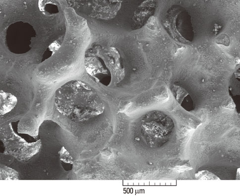
Figure 1 Scanning electron microscope image of Osteon.
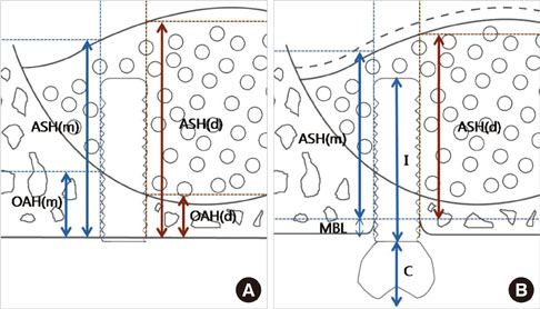
Figure 2 Schematic drawing illustrating the linear measurements taken from radiographs. (A) Immediately after the sinus augmentation. (B) 1-year after the sinus augmentation. ASH (m): mesial augmented sinus height, ASH (d): distal augmented sinus height, OAH (m): mesial original alveolar bone height, OAH (d): distal original alveolar bone height, MBL: marginal bone loss, I: implant fixture length, C: crown length.
Reference
-
1. Chiapasco M, Zaniboni M, Rimondini L. Dental implants placed in grafted maxillary sinuses: a retrospective analysis of clinical outcome according to the initial clinical situation and a proposal of defect classification. Clin Oral Implants Res. 2008. 19:416–428.
Article2. Jensen OT, Shulman LB, Block MS, Iacono VJ. Report of the Sinus Consensus Conference of 1996. Int J Oral Maxillofac Implants. 1998. 13:Suppl. 11–45.3. Pjetursson BE, Tan WC, Zwahlen M, Lang NP. A systematic review of the success of sinus floor elevation and survival of implants inserted in combination with sinus floor elevation. J Clin Periodontol. 2008. 35:8 Suppl. 216–240.
Article4. Wallace SS, Froum SJ. Effect of maxillary sinus augmentation on the survival of endosseous dental implants. A systematic review. Ann Periodontol. 2003. 8:328–343.
Article5. Hallman M, Hedin M, Sennerby L, Lundgren S. A prospective 1-year clinical and radiographic study of implants placed after maxillary sinus floor augmentation with bovine hydroxyapatite and autogenous bone. J Oral Maxillofac Surg. 2002. 60:277–284.
Article6. Johansson B, Grepe A, Wannfors K, Hirsch JM. A clinical study of changes in the volume of bone grafts in the atrophic maxilla. Dentomaxillofac Radiol. 2001. 30:157–161.
Article7. Misch CE, Dietsh F. Bone-grafting materials in implant dentistry. Implant Dent. 1993. 2:158–167.
Article8. Dalkýz M, Ozcan A, Yapar M, Gökay N, Yüncü M. Evaluation of the effects of different biomaterials on bone defects. Implant Dent. 2000. 9:226–235.
Article9. Kim YK, Yun PY, Kim SG, Lim SC. Analysis of the healing process in sinus bone grafting using various grafting materials. Oral Surg Oral Med Oral Pathol Oral Radiol Endod. 2009. 107:204–211.
Article10. Daculsi G, LeGeros RZ, Nery E, Lynch K, Kerebel B. Transformation of biphasic calcium phosphate ceramics in vivo: ultrastructural and physicochemical characterization. J Biomed Mater Res. 1989. 23:883–894.
Article11. Gauthier O, Bouler JM, Aguado E, Pilet P, Daculsi G. Macroporous biphasic calcium phosphate ceramics: influence of macropore diameter and macroporosity percentage on bone ingrowth. Biomaterials. 1998. 19:133–139.
Article12. Karabuda C, Ozdemir O, Tosun T, Anil A, Olgaç V. Histological and clinical evaluation of 3 different grafting materials for sinus lifting procedure based on 8 cases. J Periodontol. 2001. 72:1436–1442.
Article13. Nery EB, LeGeros RZ, Lynch KL, Lee K. Tissue response to biphasic calcium phosphate ceramic with different ratios of HA/beta TCP in periodontal osseous defects. J Periodontol. 1992. 63:729–735.
Article14. Yamada S, Heymann D, Bouler JM, Daculsi G. Osteoclastic resorption of biphasic calcium phosphate ceramic in vitro. J Biomed Mater Res. 1997. 37:346–352.15. Yamada S, Heymann D, Bouler JM, Daculsi G. Osteoclastic resorption of calcium phosphate ceramics with different hydroxyapatite/beta-tricalcium phosphate ratios. Biomaterials. 1997. 18:1037–1041.
Article16. Kim YK, Yun PY, Lim SC, Kim SG, Lee HJ, Ong JL. Clinical evaluations of OSTEON as a new alloplastic material in sinus bone grafting and its effect on bone healing. J Biomed Mater Res B Appl Biomater. 2008. 86:270–277.
Article17. Lee JH, Jung UW, Kim CS, Choi SH, Cho KS. Maxillary sinus augmentation using macroporous biphasic calcium phosphate (MBCP(TM)): three case report with histologic evaluation. J Korean Acad Periodontol. 2006. 36:567–577.
Article18. Zitzmann NU, Schärer P. Sinus elevation procedures in the resorbed posterior maxilla. Comparison of the crestal and lateral approaches. Oral Surg Oral Med Oral Pathol Oral Radiol Endod. 1998. 85:8–17.19. Zarb GA, Zarb FL. Tissue integrated dental prostheses. Quintessence Int. 1985. 16:39–42.20. Boyne PJ, James RA. Grafting of the maxillary sinus floor with autogenous marrow and bone. J Oral Surg. 1980. 38:613–616.21. Kent JN, Block MS. Simultaneous maxillary sinus floor bone grafting and placement of hydroxylapatite-coated implants. J Oral Maxillofac Surg. 1989. 47:238–242.
Article22. Cutler SJ, Ederer F. Maximum utilization of the life table method in analyzing survival. J Chronic Dis. 1958. 8:699–712.
Article23. Buser D, Weber HP, Lang NP. Tissue integration of non-submerged implants. 1-year results of a prospective study with 100 ITI hollow-cylinder and hollow-screw implants. Clin Oral Implants Res. 1990. 1:33–40.
Article24. Block MS, Kent JN, Kallukaran FU, Thunthy K, Weinberg R. Bone maintenance 5 to 10 years after sinus grafting. J Oral Maxillofac Surg. 1998. 56:706–714.
Article25. Chanavaz M. Maxillary sinus: anatomy, physiology, surgery, and bone grafting related to implantology--eleven years of surgical experience (1979-1990). J Oral Implantol. 1990. 16:199–209.26. Chanavaz M, Francke JP, Donazzan M. The maxillary sinus and implantology. Chir Dent Fr. 1990. 60:45–54.27. Hatano N, Shimizu Y, Ooya K. A clinical long-term radiographic evaluation of graft height changes after maxillary sinus floor augmentation with a 2:1 autogenous bone/xenograft mixture and simultaneous placement of dental implants. Clin Oral Implants Res. 2004. 15:339–345.
Article28. Maiorana C, Sigurtà D, Mirandola A, Garlini G, Santoro F. Sinus elevation with alloplasts or xenogenic materials and implants: an up-to-4-year clinical and radiologic follow-up. Int J Oral Maxillofac Implants. 2006. 21:426–432.29. Hieu PD, Chung JH, Yim SB, Hong KS. A radiographical study on the changes in height of grafting materials after sinus lift: a comparison between two types of xenogenic materials. J Periodontal Implant Sci. 2010. 40:25–32.
Article30. Kahnberg KE, Ekestubbe A, Gröndahl K, Nilsson P, Hirsch JM. Sinus lifting procedure. I. One-stage surgery with bone transplant and implants. Clin Oral Implants Res. 2001. 12:479–487.31. Keller EE, Eckert SE, Tolman DE. Maxillary antral and nasal one-stage inlay composite bone graft: preliminary report on 30 recipient sites. J Oral Maxillofac Surg. 1994. 52:438–447.
Article32. Gray CF, Redpath TW, Bainton R, Smith FW. Magnetic resonance imaging assessment of a sinus lift operation using reoxidised cellulose (Surgicel) as graft material. Clin Oral Implants Res. 2001. 12:526–530.
Article
- Full Text Links
-
- Actions
-
Cited
- CITED
-
- Close
- Share
-
- Similar articles
-
- Maxillary Sinus Augmentation Using Macroporous Biphasic Calcium Phosphate (MBCP(TM)): Three Case Report With Histologic Evaluation
- Maxillary sinus augmentation using biphasic calcium phosphate: dimensional stability results after 3–6 years
- Management of Perioperative Pathologic Conditions Involving Maxillary Sinus for Dental Implant Placement and Sinus Augmentation: Report of Case Series and Literature Review
- Increased osteoinductivity and mineralization by minimal concentration of bone morphogenetic protein-2 loaded onto biphasic calcium phosphate in a rabbit sinus
- Delayed Occurrence of Maxillary Sinusitis after Simultaneous Maxillary Sinus Augmentation and Implant: A Case Report and Literature Review
