Maxillary sinus augmentation using biphasic calcium phosphate: dimensional stability results after 3–6 years
- Affiliations
-
- 1Department of Periodontology, Research Institute for Periodontal Regeneration, Yonsei University College of Dentistry, Seoul, Korea. shchoi726@yuhs.ac
- KMID: 2440177
- DOI: http://doi.org/10.5051/jpis.2019.49.1.47
Abstract
- PURPOSE
This study was designed to observe the resorption pattern of biphasic calcium phosphate (BCP) used for maxillary sinus augmentation over a 3- to 6-year healing period, and to investigate factors affecting the resorption of BCP.
METHODS
A total of 47 implants placed in 27 sinuses of 22 patients were investigated. All patients had residual bone height less than 5 mm at baseline. The modified Caldwell-Luc approach was used to elevate the maxillary sinus membrane, and the sinus cavity was filled with BCP (70% hydroxyapatite and 30% β-tricalcium phosphate). Implant placement was done simultaneously or in a staged manner. Serial radiographic analysis was performed up to 6 years postoperatively.
RESULTS
During the follow-up period, no implant loss was reported. The mean reduced height of the augmented sinus (RHO) was 0.27±1.08 mm at 36 months, and 0.89±1.39 mm at 72 months postoperatively. Large amounts of graft material (P=0.021) and a long healing period (P=0.035) significantly influenced the amount of RHO. In particular, there was a significant relationship between a healing period longer than 40 months and RHO.
CONCLUSIONS
BCP can achieve proper dimensional stability with minimal reduction of the graft height in a 3- to 6-year healing period after maxillary sinus augmentation. The healing period and the amount of graft material influenced the resorption of BCP.
MeSH Terms
Figure
-
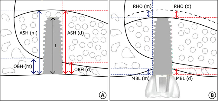
Figure 1 Schematic drawing illustrating the linear measurements taken from radiographs. (A) Immediately after the sinus augmentation. (B) Follow-up after the sinus augmentation. Figure 1 is modified from a figure in our previous study [15]. ASH(m): mesial augmented sinus height, ASH(d): distal augmented sinus height, OBH(m): mesial original alveolar bone height, OBH(d): distal original alveolar bone height, RHO(m): mesial reduced height of augmented sinus, RHO(d): distal reduced height of augmented sinus, MBL(m): mesial marginal bone loss, MBL(d): distal marginal bone loss, I: implant fixture length.
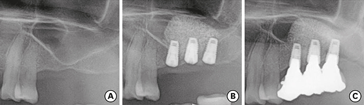
Figure 2 Panoramic radiographs. (A) Before maxillary sinus augmentation surgery. (B) Immediately after maxillary sinus augmentation and implantation surgery (1-stage). (C) Five years after surgery.
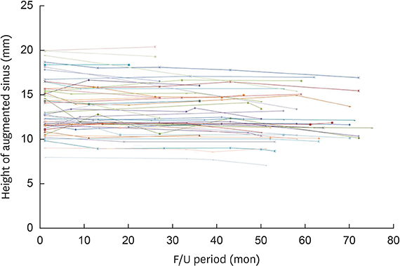
Figure 3 Graph showing changes of augmented sinus height over the follow-up period in all participants.
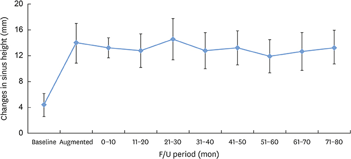
Figure 4 Mean changes of augmented sinus height over the follow-up period.
Reference
-
1. Smiler DG, Johnson PW, Lozada JL, Misch C, Rosenlicht JL, Tatum OH Jr, et al. Sinus lift grafts and endosseous implants. Treatment of the atrophic posterior maxilla. Dent Clin North Am. 1992; 36:151–186.2. Boyne PJ, James RA. Grafting of the maxillary sinus floor with autogenous marrow and bone. J Oral Surg. 1980; 38:613–616.3. Tatum H Jr. Maxillary and sinus implant reconstructions. Dent Clin North Am. 1986; 30:207–229.4. Misch CE. Maxillary sinus augmentation for endosteal implants: organized alternative treatment plans. Int J Oral Implantol. 1987; 4:49–58.5. Buchmann R, Khoury F, Faust C, Lange DE. Peri-implant conditions in periodontally compromised patients following maxillary sinus augmentation. A long-term post-therapy trial. Clin Oral Implants Res. 1999; 10:103–110.
Article6. Mohan N, Wolf J, Dym H. Maxillary sinus augmentation. Dent Clin North Am. 2015; 59:375–388.
Article7. Erbe EM, Marx JG, Clineff TD, Bellincampi LD. Potential of an ultraporous β-tricalcium phosphate synthetic cancellous bone void filler and bone marrow aspirate composite graft. Eur Spine J. 2001; 10:Suppl 2. S141–S146.
Article8. Wu J, Li B, Lin X. Histological outcomes of sinus augmentation for dental implants with calcium phosphate or deproteinized bovine bone: a systematic review and meta-analysis. Int J Oral Maxillofac Surg. 2016; 45:1471–1477.
Article9. Lambert F, Léonard A, Drion P, Sourice S, Layrolle P, Rompen E. Influence of space-filling materials in subantral bone augmentation: blood clot vs. autogenous bone chips vs. bovine hydroxyapatite. Clin Oral Implants Res. 2011; 22:538–545.
Article10. Kirmeier R, Payer M, Wehrschuetz M, Jakse N, Platzer S, Lorenzoni M. Evaluation of three-dimensional changes after sinus floor augmentation with different grafting materials. Clin Oral Implants Res. 2008; 19:366–372.
Article11. Lim HC, Hong JY, Lee JS, Jung UW, Choi SH. Late-term healing in an augmented sinus with different ratios of biphasic calcium phosphate: a pilot study using a rabbit sinus model. J Periodontal Implant Sci. 2016; 46:57–69.
Article12. Ohe JY, Kim GT, Lee JW, Al Nawas B, Jung J, Kwon YD. Volume stability of hydroxyapatite and β-tricalcium phosphate biphasic bone graft material in maxillary sinus floor elevation: a radiographic study using 3D cone beam computed tomography. Clin Oral Implants Res. 2016; 27:348–353.
Article13. Shanbhag S, Shanbhag V, Stavropoulos A. Volume changes of maxillary sinus augmentations over time: a systematic review. Int J Oral Maxillofac Implants. 2014; 29:881–892.
Article14. Kühl S, Payer M, Kirmeier R, Wildburger A, Acham S, Jakse N. The influence of particulated autogenous bone on the early volume stability of maxillary sinus grafts with biphasic calcium phosphate: a randomized clinical trial. Clin Implant Dent Relat Res. 2015; 17:173–178.
Article15. Cha JK, Park JC, Jung UW, Kim CS, Cho KS, Choi SH. Case series of maxillary sinus augmentation with biphasic calcium phosphate: a clinical and radiographic study. J Periodontal Implant Sci. 2011; 41:98–104.
Article16. Zitzmann NU, Schärer P. Sinus elevation procedures in the resorbed posterior maxilla. Comparison of the crestal and lateral approaches. Oral Surg Oral Med Oral Pathol Oral Radiol Endod. 1998; 85:8–17.17. Moraschini V, Uzeda MG, Sartoretto SC, Calasans-Maia MD. Maxillary sinus floor elevation with simultaneous implant placement without grafting materials: a systematic review and meta-analysis. Int J Oral Maxillofac Surg. 2017; 46:636–647.
Article18. Zijderveld SA, Schulten EA, Aartman IH, ten Bruggenkate CM. Long-term changes in graft height after maxillary sinus floor elevation with different grafting materials: radiographic evaluation with a minimum follow-up of 4.5 years. Clin Oral Implants Res. 2009; 20:691–700.
Article19. Cordaro L, Bosshardt DD, Palattella P, Rao W, Serino G, Chiapasco M. Maxillary sinus grafting with Bio-Oss or Straumann Bone Ceramic: histomorphometric results from a randomized controlled multicenter clinical trial. Clin Oral Implants Res. 2008; 19:796–803.
Article20. Sartori S, Silvestri M, Forni F, Icaro Cornaglia A, Tesei P, Cattaneo V. Ten-year follow-up in a maxillary sinus augmentation using anorganic bovine bone (Bio-Oss). A case report with histomorphometric evaluation. Clin Oral Implants Res. 2003; 14:369–372.
Article21. Mordenfeld A, Hallman M, Johansson CB, Albrektsson T. Histological and histomorphometrical analyses of biopsies harvested 11 years after maxillary sinus floor augmentation with deproteinized bovine and autogenous bone. Clin Oral Implants Res. 2010; 21:961–970.
Article22. Iezzi G, Scarano A, Mangano C, Cirotti B, Piattelli A. Histologic results from a human implant retrieved due to fracture 5 years after insertion in a sinus augmented with anorganic bovine bone. J Periodontol. 2008; 79:192–198.
Article23. Lee JS, Cha JK, Thoma DS, Jung UW. Report of a human autopsy case in maxillary sinuses augmented using a synthetic bone substitute: micro-computed tomographic and histologic observations. Clin Oral Implants Res. 2018; 29:339–345.
Article24. Mordenfeld A, Lindgren C, Hallman M. Sinus floor augmentation using Straumann® BoneCeramic™ and Bio-Oss® in a split mouth design and later placement of implants: a 5-year report from a longitudinal study. Clin Implant Dent Relat Res. 2016; 18:926–936.
Article25. Okada T, Kanai T, Tachikawa N, Munakata M, Kasugai S. Long-term radiographic assessment of maxillary sinus floor augmentation using beta-tricalcium phosphate: analysis by cone-beam computed tomography. Int J Implant Dent. 2016; 2:8.
Article26. Hatano N, Shimizu Y, Ooya K. A clinical long-term radiographic evaluation of graft height changes after maxillary sinus floor augmentation with a 2:1 autogenous bone/xenograft mixture and simultaneous placement of dental implants. Clin Oral Implants Res. 2004; 15:339–345.
Article27. Wiltfang J, Schultze-Mosgau S, Nkenke E, Thorwarth M, Neukam FW, Schlegel KA. Onlay augmentation versus sinuslift procedure in the treatment of the severely resorbed maxilla: a 5-year comparative longitudinal study. Int J Oral Maxillofac Surg. 2005; 34:885–889.
Article28. Laurell L, Lundgren D. Marginal bone level changes at dental implants after 5 years in function: a meta-analysis. Clin Implant Dent Relat Res. 2011; 13:19–28.
Article
- Full Text Links
-
- Actions
-
Cited
- CITED
-
- Close
- Share
-
- Similar articles
-
- Maxillary Sinus Augmentation Using Macroporous Biphasic Calcium Phosphate (MBCP(TM)): Three Case Report With Histologic Evaluation
- Late-term healing in an augmented sinus with different ratios of biphasic calcium phosphate: a pilot study using a rabbit sinus model
- Combined Application of Dentin Noncollagenous Proteins and Odontogenic Biphasic Calcium Phosphate in Rabbit Maxillary Sinus Lifting
- Increased osteoinductivity and mineralization by minimal concentration of bone morphogenetic protein-2 loaded onto biphasic calcium phosphate in a rabbit sinus
- Case series of maxillary sinus augmentation with biphasic calcium phosphate: a clinical and radiographic study
