Nat Prod Sci.
2016 Jun;22(2):140-145. 10.20307/nps.2016.22.2.140.
Phenolic Constituents and Their Anti-inflammatory Activity from Echinochloa utilis Grains
- Affiliations
-
- 1College of Pharmacy, Catholic University of Daegu, Gyeongsan 38430, Republic of Korea. woomh@cu.ac.kr
- 2Laboratory of Immunobiology, School of Life Science and Biotechnology, College of Natural Sciences, Kyungpook National University, Daegu 39061, Republic of Korea.
- 3Functional Cereal Crop research Division, Department of Functional Crop, NICS, RDA, 50426, Republic of Korea.
- 4Phu Tho College of Pharmacy, Viettri City, Phutho Province 290000, Vietnam.
- KMID: 2328881
- DOI: http://doi.org/10.20307/nps.2016.22.2.140
Abstract
- Seven phenolic compounds including p-coumaric acid (1), 4-hydroxybenzoic acid (2), 4-hydroxybenzaldehyde (3), vanillic acid (4), luteolin (5), acacetin (6), and tricin (7), were isolated from the methylene chloride and ethyl acetate fractions of Echinochloa utilis grains. Compounds (1 - 4, 6) were isolated for the first time from this plant. These compounds were tested for inhibitory activities against LPS-induced NO production in RAW 264.7 cells. Compounds 5 and 6 displayed significant inhibitory effects, with ICâ‚…â‚€ values of 27.9 ± 2.6 and 14.0 ± 1.1 µM, respectively. The results suggested that E. utilis ethanolic extract may be used as a potential source of anti-inflammatory agents and functional foods for the treatment of allergic diseases.
MeSH Terms
Figure
-
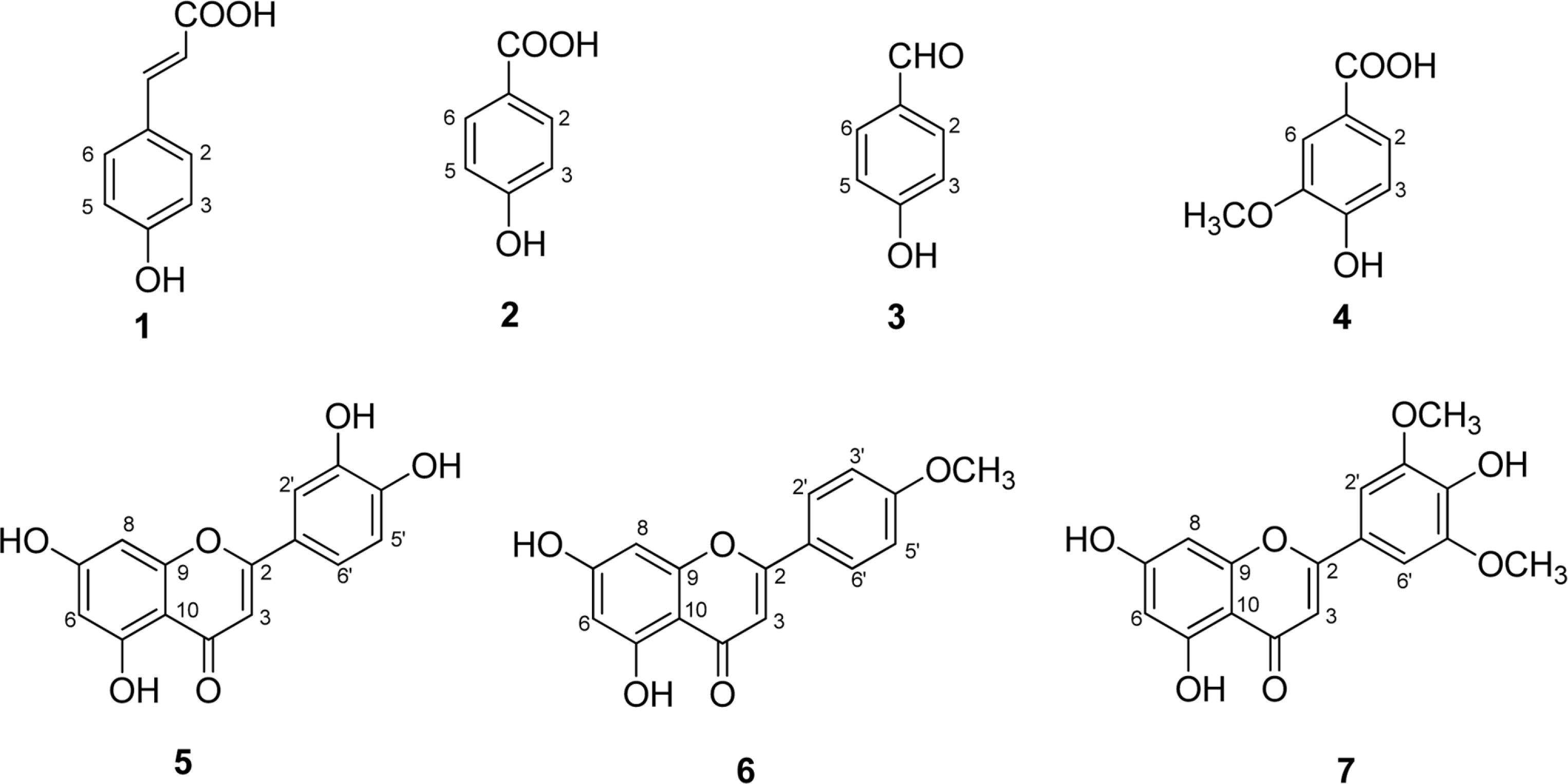
Fig. 1. Chemical structures of compounds 1–7 isolated from the grains of E. utilis.

Fig. 2. The effects of compounds 5 and 6 on LPS-induced rise of iNOS and COX-2 levels in RAW 264.7 cells. The RAW 264.7 cells were pretreated with LPS (0.1 µg/mL) for 30 min, followed by concentrations of 2.5, 5.0, and 10.0 µg/mL for 5 and 1.25, 2.5, and 5.0 µg/mL for 6 for 24 h. Whole-cell lysates were blotted with the indicated antibodies. β-Actin level was used as a loading control. The expressions of iNOS and COX-2 were assessed by Western blotting analysis using specific antibodies for individual proteins. The results are representatives of three independent experiments.
Reference
-
(1). Zedler S., Faist E.Curr. Opin. Crit. Care. 2006; 12:595–601.(2). Mariathasan S., Monack D. M.Nat. Rev. Immunol. 2007; 7:31–40.(3). Korhonen R., Lahti A., Kankaanranta H., Moilanen E.Curr. Drug Targets Inflamm. Allergy. 2005; 4:471–479.(4). Salvemini D., Ischiropoulos H., Cuzzocrea S.Methods Mol. Biol. 2003; 225:291–303.(5). Yabuno T.Econ. Bot. 1987; 41:484–493.(6). Nozawa S., Takahashi M., Nakai H., Sato Y.Breed. Sci. 2006; 56:335–340.(7). Park K.Y., Park R. K., Choi B. H.Korean J. Crop Sci. 1991; 36:249–253.(8). Kim J. Y., Jang K. C., Park B. R., Han S. I., Choi K. J., Kim S. Y., Oh S. H., Ra J. E., Ha T. J., Lee J. H., Hwang J. Y., Kang H. W., Seo W. D.Food Sci. Biotechnol. 2011; 20:461–469.(9). Watanabe M. J.Agric. Food Chem. 1999; 47:4500–4505.(10). Migliorini P., Corradin G., Corradin S. B. J.Immunol. Methods. 1991; 139:107–114.(11). Lee Y. J., Han J. Y., Lee C. G., Heo K., Park S. I., Park Y. S., Kim J. S., Yang K. M., Lee K. J., Kim T. H., Rhee M. H., Kim S. D. J.Ginseng Res. 2014; 38:208–214.(12). Yang J., Wang D., Liu W., Zhang X., Bian F., Yu W.Green Chem. 2013; 15:3429–3437.(13). Yayli N., Yildirim N., Usta A., Özkurt S., Akgün V., Turk J.Chem. 2003; 27:749–756.(14). Xu M. L., Wang L., Hu J. H., Wang M. H. J.Food Sci. Nutr. 2009; 14:354–357.(15). Chang S. W., Kim K. H., Lee I. K., Choi S. U., Ryu S. Y., Lee K. R.Nat. Prod. Sci. 2009; 15:234–240.(16). Wagner H., Chari V. M., Sonnenbichler J.Tetrahedron Lett. 1976; 17:1799–1802.(17). Shimoi K., Masuda S., Furugori M., Esaki S., Kinae N.Carcinogenesis. 1994; 15:2669–2672.(18). Yamamoto H., Sakakibara J., Nagatsu A., Sekiya K. J.Agric. Food Chem. 1998; 46:862–865.(19). Lin Y., Shi R., Wang X., Shen H. M.Curr. Cancer Drug Targets. 2008; 8:634–646.(20). Seelinger G., Merfort I., Schempp C. M.Planta Med. 2008; 74:1667–1677.(21). Park C. M., Song Y. S.Nutr. Res. Pract. 2013; 7:423–429.(22). Yang Z. G., Jia L. N., Shen Y., Ohmura A., Kitanaka S.Molecules. 2011; 16:8305–8318.(23). Chan T. S., Galati G., Pannala A. S., Rice-Evans C., O'Brien P. J.Free Radic. Res. 2003; 37:787–794.(24). Pan M. H., Lai C. S., Wang Y. J., Ho C. T.Biochem. Pharmacol. 2006; 72:1293–1303.(25). Srisook K., Srisook E., Nachaiyo W., Chan-In M., Thongbai J., Wongyoo K., Chawsuanthong S., Wannasri K., Intasuwan S., Watcharanawee K. J.Ethnopharmacol. 2015; 165:94–102.
- Full Text Links
-
- Actions
-
Cited
- CITED
-
- Close
- Share
-
- Similar articles
-
- Preparation and Evaluation of Anti-Emetic Ginger Orodispersible Tablets from Standardized Extract using Phenolic Profile Effects
- Analysis of Phenolic Acid Content and Antioxidant Activity of Chestnut Honey from Different Regions of Korea
- Antioxidant Activity and Phenolic Content of Different Parts of Lotus and Optimization of Extraction Condition using Response Surface Methodology
- Phenolic Constituents from Balanophora laxiflora with their Anti-inflammatory and Cytotoxic Effects
- Biological Activity of Chemical Constituents Isolated from Strain Chlamydomonas sp. KSF108 (Chlamydomonadaceae)
