J Korean Assoc Oral Maxillofac Surg.
2010 Oct;36(5):417-422.
The study of bone density assessment on dental implant sites
- Affiliations
-
- 1Department of Oral and Maxillofacial Surgery, Ulsan University Hospital, College of Medicine, University of Ulsan, Ulsan, Korea. lovenip@mail.ulsan.ac.kr
- 2Department of Occupational and Environmental Medicine, College of Medicine, University of Ulsan, Ulsan, Korea.
Abstract
- INTRODUCTION
Bone density is one of the important factors for the long term success of endosseous implants. The bone density varies from site to site and from patient to patient. A preoperative evaluation of the bone density is quite useful to oral surgeons for planning dental implantation. More accurate information on the bone density will help surgeons identify suitable implant sites, thereby increase the success rate of dental implantation. This study examined the correlation between the bone density measured preoperatively by computed tomography (CT) and the implant primary stability measured by resonance frequency analysis. Furthermore, the effects of the implant sites, gender, age and generalized systemic disorder patients on the bone density and primary implant stability were examined.
MATERIALS AND METHODS
One hundred and fourteen patients were selected. None of the patients had undergone a tooth extraction or bone graft history in the previous year. Preoperatively, the patients underwent CT scanning to evaluate the Hounsfield unit (HU), and resonance frequency analysis (RFA) was used to evaluate the implant primary stability at the time of implant installation. All implants were 4.0 mm diameter and 11.5 mm length US II. All patients were recorded and the HU and implant stability quotient (ISQ) value were evaluated according to the sites, gender and age.
RESULTS
The highest HU values were found in the mandibular anterior site (827.6+/-151.4), followed by the mandibular molar site (797+/-135.1), mandibular premolar site (753.8+/-171.2), maxillary anterior site (726.3+/-154.4), maxillary premolar site (656.7+/-173.8) and maxillary molar site (621.5+/-164.9). The ISQ value was the highest in the mandibular premolar site (81.5+/-2.4) followed by the mandibular molar site (80.0+/-5.7), maxillary anterior site (77.4+/-4.1), mandibular anterior site (76.4+/-11.9), maxillary premolar site (74.2+/-14.3) and maxillary molar site (73.7+/-7.4). The mean HU and ISQ value were similar in females and males. (HU: P=0.331, ISQ: P=0.595) No significant difference was also found in the age group respectively. However, the correlation coefficients between the variables showed a closed correlation between the HU and ISQ value.
CONCLUSION
These results showed close correlation between the bone density (HU) and primary stability value (ISQ) at the time of implant installation (Correlation coefficients=0.497, P<0.01). These results strengthen the hypothesis that it might be possible to predict and quantify the initial implant stability and bone density from a presurgical CT diagnosis. These results strengthen the hypothesis that it might be possible to predict and quantify the initial implant stability and bone density from a presurgical CT diagnosis.
Keyword
MeSH Terms
Figure
-

Fig. 1. Image of transaxial cut of Somatom computed tomography (CT). The hounsfield unit (HU) measurement feature of CT was utilized to evaluate the bone density.
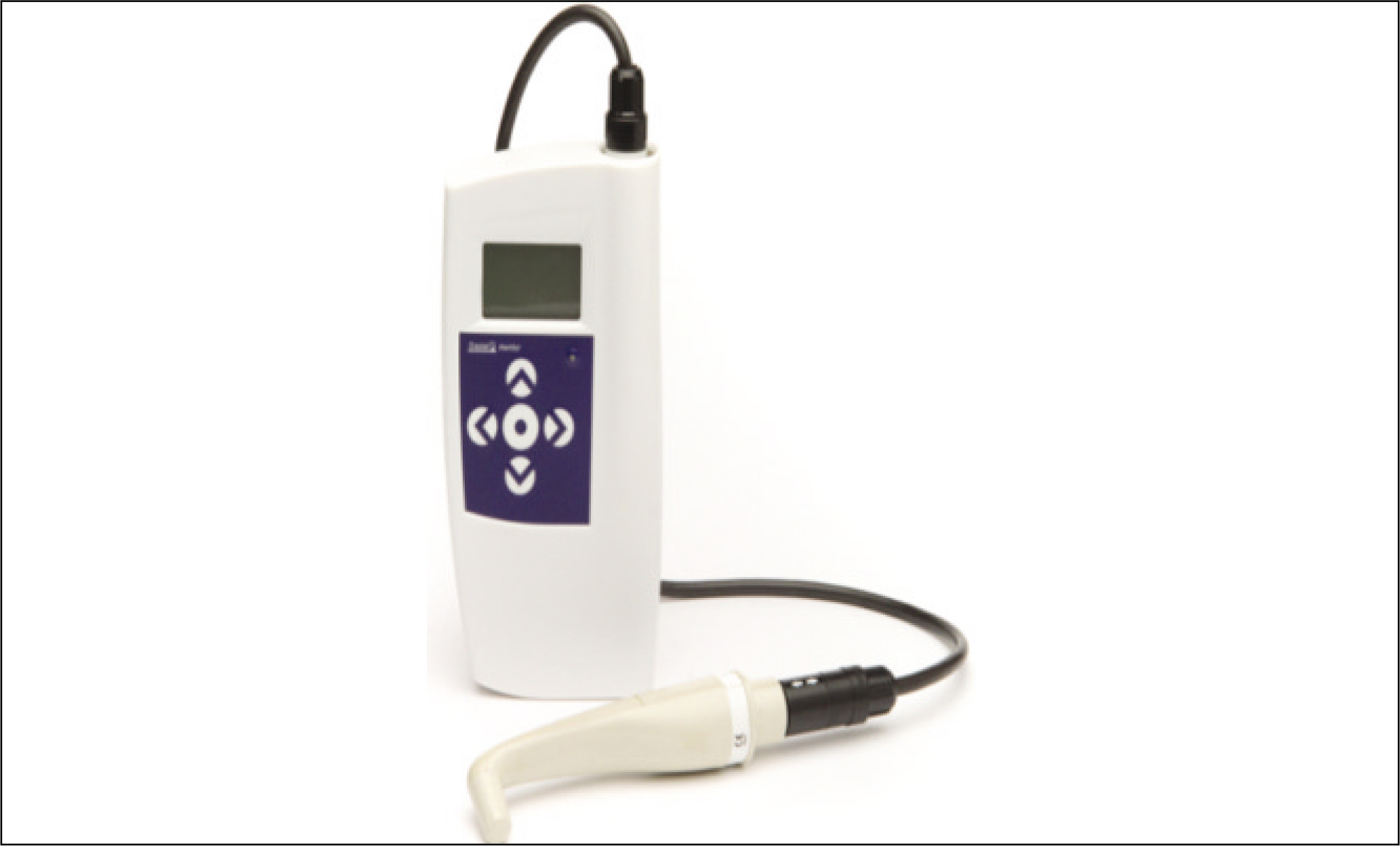
Fig. 2. Osstell mentor to measure implant stability quotient (ISQ) value.
Reference
-
References
1. Turkyilmaz I, To ¨zu ¨m TF, Tumer C. Bone density assessments of oral implant sites using computerized tomograpy. J Oral Rehabil. 2007; 34:267–72.2. de Oliveira RC, Leles CR, Normanha LM, Lindh C, Ribeiro-Rotta RF. Assessments of trabecular bone density at implant sites on CT images. Oral Surg Oral Med Oral Pathol Oral Radiol Endod. 2008; 105:231–8.
Article3. Lekholm U, Zarb GA. Patient selection and preparation. Bra�nemark PI, Zarb GA, Albrektsson T, editors. Tissue integrated prostheses: osseointegration in clinical dentistry. Chicago: Quintessence;1985. p. 199–209.4. Misch C. Classifications and treatment options of the completely edentulous arch in implant dentistry. Dent Today. 1990; 9(26):28–30.5. Norton MR, Gamble C. Bone classification: an objective scale of bone density using the computerized tomography scan. Clin Oral Implants Res. 2001; 12:79–84.
Article6. Hounsfield GN. Computerized transverse axial scanning (tomography): Part I. Description of system 1973. Br J Radiol. 1995; 68:H166–72.7. Fanuscu MI, Chang TL. Three-dimensional morphometric analysis of human cadaver bone: microstructural data from maxilla and mandible. Clin Oral Implants Res. 2004; 15:213–8.
Article8. Martinez H, Davarpanah M, Missika P, Celletti R, Lazzara R. Optimal implant stabilization in low density bone. Clin Oral Implants Res. 2001; 12:423–32.
Article9. Meredith N, Alleyne D, Cawley P. Quantitative determination of the stability of the implant-tissue interface using resonance frequency analysis. Clin Oral Implants Res. 1996; 7:261–7.
Article10. Turkyilmaz I, Tumer C, Ozbek EN, To ¨zu ¨m TF. Relations between the bone density values from computerized tomography, and implant stability parameters: a clinical study of 230 regular platform implants. J Clin Periodontol. 2007; 34:716–22.
Article11. Turkyilmaz I, Tumer C, Ozbek EN, To ¨zu ¨m TF. Assessment of correlation between computerized tomography values of bone, and maximum torque and resonance frequency values at dental implant placement. J Oral Rehabil. 2006; 33:881–8.12. Sennerby L, Meredith N. Resonance frequency analysis: measuring implant stability and osseointegration. Compend Contin Educ Dent. 1998; 19:493–8. 500, 502; quiz 504.13. Shapurian T, Damoulis PD, Reiser GM, Griffin TJ, Rand WM. Quantitative evaluation of bone density using the Hounsfield index. Int J Oral Maxillofac Implants. 2006; 21:290–7.14. Shahlaie M, Gantes B, Schulz E, Riggs M, Crigger M. Bone density assessments of dental implant sites: 1. Quantitative computed tomography. Int J Oral Maxillofac Implants. 2003; 18:224–31.15. Park HS, Lee YJ, Jeong SH, Kwon TG. Density of the alveolar and basal bones of the maxilla and the mandible. Am J Orthod Dentofacial Orthop. 2008; 133:30–7.
Article16. Bassi F, Procchio M, Fava C, Schierano G, Preti G. Bone density in human dentate and edentulous mandible using computed tomography. Clin Oral Implants Res. 1999; 10:356–61.17. Lee S, Gantes B, Riggs M, Crigger M. Bone density assessments of dental implant sites: 3. Bone quality evaluation during osteotomy and implant placement. Int J Oral Maxillofac Implants. 2007; 22:208–12.18. Fanfani F, Pierazzini A. La tomografia assilae computerizzata del distetto maxillo-facciale, 3D-Dentascan e derivati. Torino, Italy: UTET Perioici;1996.19. Ostman PO, Hellman M, Wendelhag I, Sennerby L. Resonance frequency analysis measurements of implants at placement surgery. Int J Prosthodont. 2006; 19:77–83. discussion 84.
- Full Text Links
-
- Actions
-
Cited
- CITED
-
- Close
- Share
-
- Similar articles
-
- Assessment of the increased calcification of the jaw bone with CT-Scan after dental implant placement
- ASSESSMENT OF BONE DENSITY ON MAXILLA AFTER IMPLANTATION WITH CONE BEAM COMPUTED TOMOGRAPHY
- The effect of undersizing and tapping on bone to implant contact and implant primary stability: A histomorphometric study on bovine ribs
- The effects of bone density and crestal cortical bone thickness on micromotion and peri-implant bone strain distribution in an immediately loaded implant: a nonlinear finite element analysis
- Observation of the change of the dental implant stability and bone density evaluation methods
