Analysis of stress distribution of tooth restored with metal-ceramic crown covering abfraction lesion according to its finish line location under occlusal load
- Affiliations
-
- 1Department of Prosthodontics, School of Dentistry,Yonsei University, Seoul, Republic of Korea. jfshim@yuhs.ac
- 2Department of Medical Device Evaluation, Cardiovascular Devices Division, Yonsei University, Seoul, Republic of Korea.
- 3Korea Food and Drug Administration, Seoul, Republic of Korea.
- KMID: 2321792
- DOI: http://doi.org/10.4047/jkap.2014.52.4.305
Abstract
- PURPOSE
When the full veneer crown was treated in the tooth with abfraction lesion due to various causes, the prognosis of it may be compromised according to the location of the finish line, but there is few study about the location of its buccal finish line. The purpose of this study was to investigate the effect of location of the finish line of the full veneer crown on stress distribution of the tooth with abfraction lesion.
MATERIALS AND METHODS
The two dimensional finite element model was developed to express tooth, surrounding tissue and full veneer crown. The stress distribution under eccentric 144 N occlusal load was analyzed using finite element analysis. The location of finish line was set just at the lower border of the lesion (Group 0), 1 mm (Group 1) and 2 mm (Group 2) below the lower border of the lesion.
RESULTS
In the Group 0, von Mises stress was concentrated at the finish line and the apex of the lesion. Also, the stress at the bucal finish line propagated to the lingual side. In the Group 1 and Group 2, stress distribution was similar each other. Stress was concentrated at the apex of lesion, but the stress at the buccal finish line did not propagate to the lingual side. That implied decrease of the possibility of horizontal crown fracture.
CONCLUSION
Full veneer crown alleviated the stress concentrated at the apex of the abfraction lesion, when the finish line of full veneer crown was set below the lower border of abfraction lesion.
Figure
-

Fig. 1. 2-dimensional normal tooth geometry.
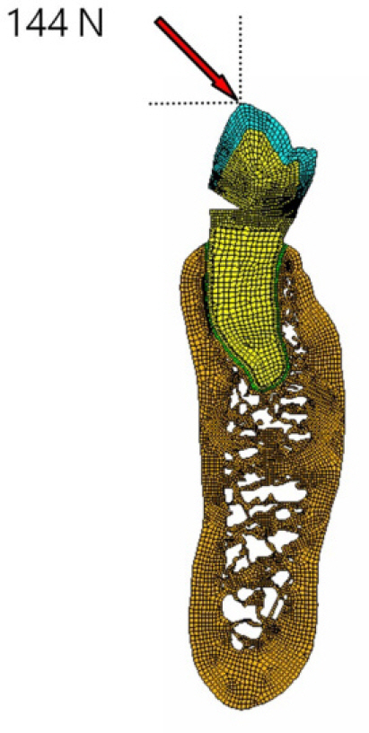
Fig. 2. Abfraction finite element model.
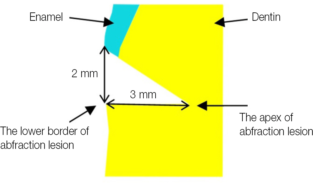
Fig. 3. Dimension of wedge-shaped defect.
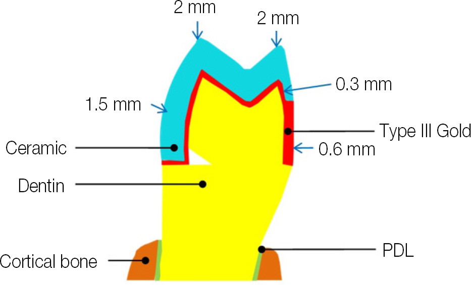
Fig. 4. Minimum recommended dimensions for a metal-ceramic restoration.
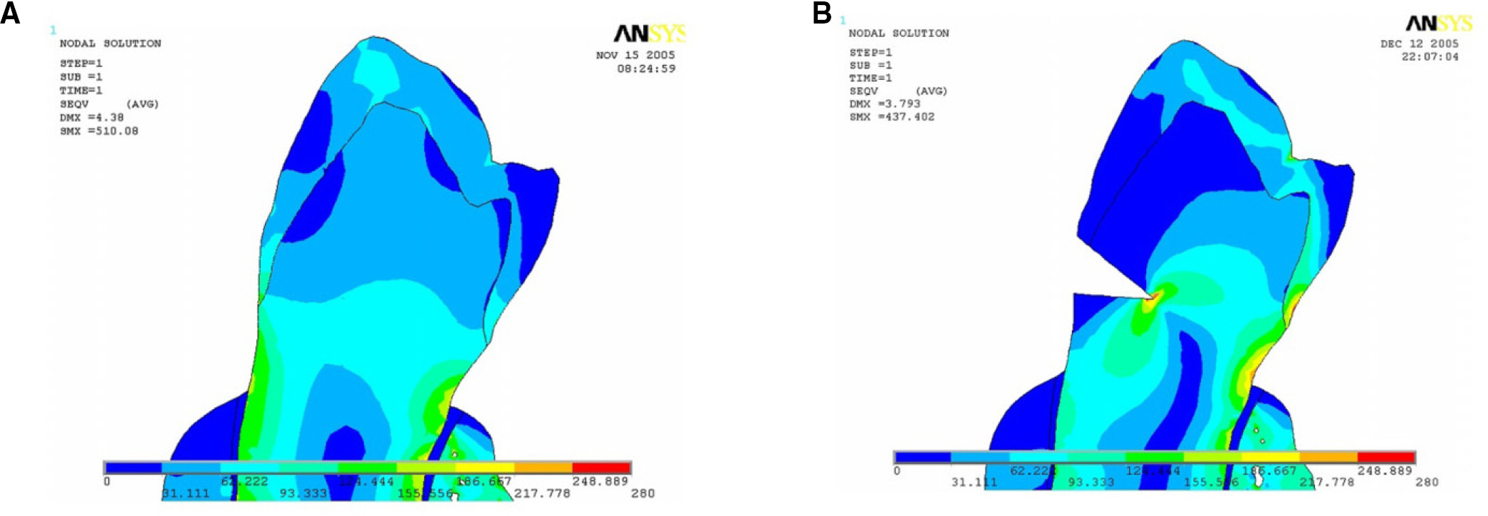
Fig. 5. von Mises stress distribution of normal tooth and the tooth with abfraction lesion. (A) Normal tooth, (B) Tooth with abfraction lesion.
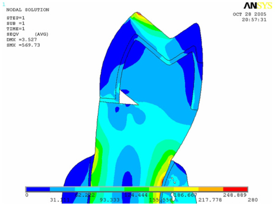
Fig. 6. von Mises stress distribution in the case that the finish line of metal-ceramiccrown is set just at the lower border of wedge-shaped defect.
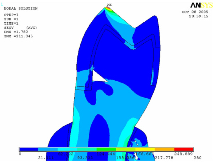
Fig. 7. von Mises stress distribution in the case that the finish line of metal-ceramic crown is set 1 mm below the lower border of wedge-shaped defect.
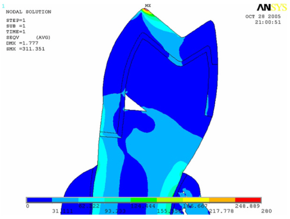
Fig. 8. von Mises stress distribution in the case that the finish line of metal-ceramic crown is set 2 mm below the lower border of wedge-shaped defect.
Reference
-
1. Grippo JO. Abfractions: a new classification of hard tissue lesions of teeth. J Esthet Dent. 1991; 3:14–9.
Article2. Lee WC, Eakle WS. Possible role of tensile stress in the etiology of cervical erosive lesions of teeth. J Prosthet Dent. 1984; 52:374–80.
Article3. Rees JS. The effect of variation in occlusal loading on the development of abfraction lesions: a finite element study. J Oral Rehabil. 2002; 29:188–93.
Article4. Francisconi LF, Graeff MS, Martins Lde M, Franco EB, Mondelli RF, Francisconi PA, Pereira JC. The effects of occlusal loading on the margins of cervical restorations. J Am Dent Assoc. 2009; 140:1275–82.
Article5. Ichim I, Schmidlin PR, Kieser JA, Swain MV. Mechanical evaluation of cervical glass-ionomer restorations: 3D finite element study. J Dent. 2007; 35:28–35.
Article6. Grippo JO. Noncarious cervical lesions: the decision to ignore or restore. J Esthet Dent. 1992; 4:55–64.
Article7. Rosenstiel SF, Land MF, Fujimoto J. Contemporary fixed prosthodontics. 3rd ed.St. Louis: Mosby;2001. xi. p. 868.8. Rubin C, Krishnamurthy N, Capilouto E, Yi H. Stress analysis of the human tooth using a three-dimensional finite element model. J Dent Res. 1983; 62:82–6.9. Rees JS, Jacobsen PH. Modelling the effects of enamel anisotropy with the finite element method. J Oral Rehabil. 1995; 22:451–4.10. Yaman SD, Alaçam T, Yaman Y. Analysis of stress distribution in a maxillary central incisor subjected to various post and core applications. J Endod. 1998; 24:107–11.
Article11. Versluis A, Douglas WH, Cross M, Sakaguchi RL. Does an in-cremental filling technique reduce polymerization shrinkage stresses? J Dent Res. 1996; 75:871–8.
Article12. Palamara D, Palamara JE, Tyas MJ, Messer HH. Strain patterns in cervical enamel of teeth subjected to occlusal loading. Dent Mater. 2000; 16:412–9.
Article13. Sorensen JA, Engelman MJ. Ferrule design and fracture resistance of endodontically treated teeth. J Prosthet Dent. 1990; 63:529–36.
Article14. Rees JS, Jacobsen PH. Elastic modulus of the periodontal liga-ment. Biomaterials. 1997; 18:995–9.
Article15. Vincent JFV. Structural biomaterials. Rev. ed.Princeton, NJ: Princeton University Press;1990. xii:p. 244.16. Lewinstein I, Banks-Sills L, Eliasi R. Finite element analysis of a new system (IL) for supporting an implant-retained cantilever prosthesis. Int J Oral Maxillofac Implants. 1995; 10:355–66.
- Full Text Links
-
- Actions
-
Cited
- CITED
-
- Close
- Share
-
- Similar articles
-
- Effect of the marginal position of prosthesis on stress distribution of teeth with abfraction lesion using finite element analysis
- THREE-DIMENSIONAL FINITE ELEMENT ANALYSIS OF STRESS DISTRIBUTION IN ALL-CERAMIC CROWNS WITH VARIOUS FINISH LINE DESIGNS AND INCISAL REDUCTIONS UNDER DIFFERENT LOADING CONDITIONS
- The fracture resistance and stress distribution of all ceramic crowns with two types of finish line on maxillary central incisor
- A study on the stress distribution of the all ceramic crowns according to the cusp angle of the maxillary first premolar using three dimensional finite element method
- Alternate metal framework designs for the metal ceramic prosthesis to enhance the esthetics
