Tuberc Respir Dis.
2009 Nov;67(5):458-461.
A Case of Pseudochylothorax Developed from Chronic Pleural Effusion after Treatment of Tuberculous Pleurisy
- Affiliations
-
- 1Department of Internal Medicine, Eulji Hospital, Eulji University School of Medicine, Seoul, Korea. hoonakr@eulji.ac.kr
- 2Department of Clinical Pathology, Eulji Hospital, Eulji University School of Medicine, Seoul, Korea.
Abstract
- A pseudochylothorax, a chyliform pleural effusion, is a rare disease of pleural effusion that contains cholesterol crystals or high lipid content that is not the result of a disrupted thoracic duct. Most of the cases were found in patients with long-standing pleural effusion due to chronic inflammatory disease, such as old tuberculous pleurisy or chronic rheumatoid pleurisy. We experienced a case of pseudochylothorax in a 74-year-old man, who was being treated for pulmonary tuberculosis and pleurisy 10 years ago. The diagnosis was confirmed on pathological study of the pleural effusion, which contained cholesterol crystals having a diagnostic rhomboid appearance.
Keyword
MeSH Terms
Figure
-
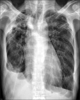
Figure 1 Simple chest X-ray demonstrates right loculated pleural effusion and bilateral pleural thickening.
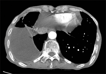
Figure 2 Chest CT scan reveals fibrotic change and multiple calcified nodules with grossly thickening of the right pleural membranes enclosing a massive effusion.

Figure 3 Microscopic examination of pleural effusion. Window-pane with a corner missing appearance of cholesterol crystals is seen in pleural effusion (×400).
Reference
-
1. Garcia-Zamalloa A, Ruiz-Irastorza G, Aguayo FJ, Gurrutxaga N. Pseudochylothorax. Report of 2 cases and review of the literature. Medicine (Baltimore). 1999. 78:200–207.2. Hillerdal G. Chylothorax and pseudochylothorax. Eur Respir J. 1997. 10:1157–1162.3. Hillerdal G. Chyliform (cholesterol) pleural effusion. Chest. 1985. 88:426–428.4. Light RW. Light RW, editor. Chapter 23. Chylothorax and pseudochylothorax. Pleural diseases. 2001. 4th ed. Philadelphia: Lippincott Williams & Wilkins;327–343.5. Zuckner J, Uddin J, Gantner GE Jr, Dorner RW. Cholesterol crystals in synovial fluid. Ann Intern Med. 1964. 60:436–446.6. Chung HR, Shon HE, Park MS. A case of cholesterol crystals in pleural effusion. J Clin Pathol Qual Control. 2000. 22:263–264.7. Coe JE, Aikawa JK. Cholesterol pleural effusion. Report of 2 cases studied with isotopic techniques and review of the world literature. Arch Intern Med. 1961. 108:763–774.8. González R, Ramírez-Rivera J. Chyliform (pseudochylous) pleural effusion. Bol Asoc Med P R. 1994. 86:50–52.9. Liss M, Brandt LJ, Wolf EL. Cholesterol crystal pseudoascites: an unusual presentation of ovarian cyst. Am J Gastroenterol. 1982. 77:245–247.10. Hamm H, Pfalzer B, Fabel H. Lipoprotein analysis in a chyliform pleural effusion: implications for pathogenesis and diagnosis. Respiration. 1991. 58:294–300.11. Song JW, Im JG, Goo JM, Kim HY, Song CS, Lee JS. Pseudochylous pleural effusion with fat-fluid levels: report of six cases. Radiology. 2000. 216:478–480.
- Full Text Links
-
- Actions
-
Cited
- CITED
-
- Close
- Share
-
- Similar articles
-
- Surgical treatment of recurrent pseudochylothorax occurring after therapy of tuberculous pleurisy
- Tuberculous Pleurisy: An Update
- Diagnostic Efficacy of Adenosine Deaminase Isoenzyme in Tuberculous Pleurisy
- A Study on pH and PCO2, in Tuberculous Pleurisy with Effusion
- Tuberculous Pleural Effusion vs Empyema: It is Possible to Differentiate Based on CT Findings?
