Restor Dent Endod.
2013 Aug;38(3):172-177.
Dilemmas pertaining to three canals in the mesiobuccal root of a maxillary second molar: a case report
- Affiliations
-
- 1Department of Conservative Dentistry and Endodontics, Manubhai Patel Dental College, Hospital and Oral Research Institute, Vadodara, India. aroraankit24@gmail.com
- 2Department of Conservative Dentistry and Endodontics, Manipal College of Dental Sciences, Manipal University, Karnataka, India.
- 3Department of Orthodontics and Dentofacial Orthopaedics, Manubhai Patel Dental College, Hospital and Oral Research Institute, Vadodara, India.
Abstract
- The mesiobuccal root of the maxillary molars is well known to pose a hindrance during endodontic therapy. Presented here is a case of a maxillary left second molar where three canals were located in its mesiobuccal root with the use of visual and diagnostic aids. Difficulties encountered during the process of unveiling the tooth's internal anatomy were discussed. The dilemmas encountered pertained to the root canal configuration, the nomenclature of the extra canals, and the justification for the presence of a third canal. The root canal configuration of 3-2-1 was confirmed for the mesiobuccal root using information gained from clinical, radiographic, and multi-detector computed tomography (MDCT) scan findings. This case demonstrates the need for efforts to locate extra canals in the mesiobuccal root of the maxillary molars as their internal anatomy remains a mystery.
Keyword
Figure
-
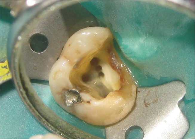
Figure 1 Three canals seen in the mesiobuccal root.
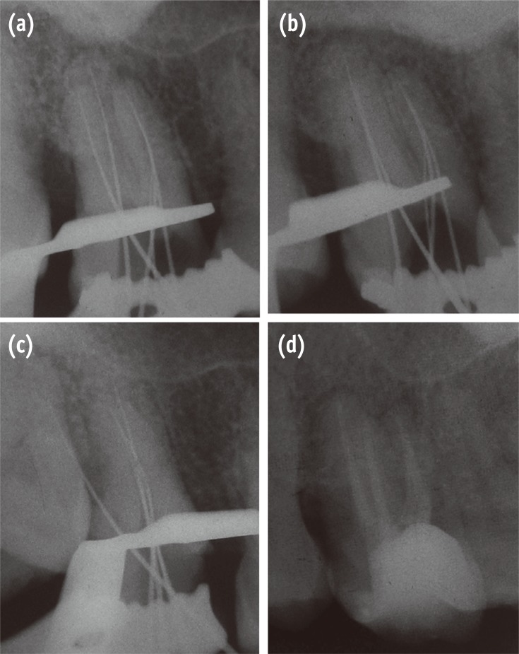
Figure 2 Radiographic findings. (a) Straight on angulation; (b) Mesial angulation; (c) Distal angulation; (d) Post obturation.
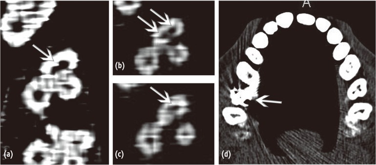
Figure 3 MDCT findings. (a) Orifice level; (b) Mid root level; (c) Apical region; (d) Source of artifact.

Figure 4 Schematic representation of canal configuration. MB, mesiobuccal; m-MB, middle mesiobuccal; p-MB, palatal mesiobuccal.
Reference
-
1. Weine FS, Healey HJ, Gerstein H, Evanson L. Canal configuration in the mesiobuccal root of the maxillary first molar and its endodontic significance. Oral Surg Oral Med Oral Pathol. 1969; 28:419–425. PMID: 5257186.
Article2. Neaverth EJ, Kotler LM, Kaltenbach RF. Clinical investigation (in vivo) of endodontically treated maxillary first molars. J Endod. 1987; 13:506–512. PMID: 3482228.3. Cheung GS. Endodontic failures - changing the approach. Int Dent J. 1996; 46:131–138. PMID: 8886865.4. Vertucci FJ. Root canal morphology and its relationship to endodontic procedures. Endod Topics. 2005; 10:3–29.
Article5. Eskoz N, Weine FS. Canal configuration of the mesiobuccal root of the maxillary second molar. J Endod. 1995; 21:38–42. PMID: 7714433.
Article6. Stropko JJ. Canal morphology of maxillary molars: clinical observations of canal configurations. J Endod. 1999; 25:446–450. PMID: 10530248.
Article7. Blattner TC, George N, Lee CC, Kumar V, Yelton CD. Efficacy of cone-beam computed tomography as a modality to accurately identify the presence of second mesiobuccal canals in maxillary first and second molars: a pilot study. J Endod. 2010; 36:867–870. PMID: 20416435.
Article8. Neelakantan P, Subbarao C, Ahuja R, Subbarao CV, Gutmann JL. Cone-beam computed tomography study of root and canal morphology of maxillary first and second molars in an Indian population. J Endod. 2010; 36:1622–1627. PMID: 20850665.
Article9. Vertucci FJ. The endodontic significance of the mesiobuccal root of the maxillary first molar. US Navy Med. 1974; 63:29–31. PMID: 4530549.10. Hess W, Zurcher E. The anatomy of the root canals of the teeth of the permanent and deciduous dentition. New York: William Wood & Co.;p. 192–195.11. Schilder H. Filling root canals in three dimensions. Dent Clin North Am. 1967; 11:723–744. PMID: 5262492.
Article12. Schilder H. Cleaning and shaping the root canal. Dent Clin North Am. 1974; 18:269–296. PMID: 4522570.13. Degerness RA, Bowles WR. Dimension, anatomy and morphology of the mesiobuccal root canal system in maxillary molars. J Endod. 2010; 36:985–989. PMID: 20478451.
Article14. Calişkan MK, Pehlivan Y, Sepetçioğlu F, Türkün M, Tuncer SS. Root canal morphology of human permanent teeth in a Turkish population. J Endod. 1995; 21:200–204. PMID: 7673821.15. Ferguson DB, Kjar KS, Hartwell GR. Three canals in the mesiobuccal root of a maxillary first molar: a case report. J Endod. 2005; 31:400–402. PMID: 15851938.
Article16. Ozcan E, Aktan AM, Ari H. A case report: unusual anatomy of maxillary second molar with 3 mesiobuccal canals. Oral Surg Oral Med Oral Pathol Oral Radiol Endod. 2009; 107:e43–e46. PMID: 19101482.
Article17. Pineda F, Kuttler Y. Mesiodistal and buccolingual roentgenographic investigation of 7,275 root canals. Oral Surg Oral Med Oral Pathol. 1972; 33:101–110. PMID: 4500261.
Article18. Vertucci FJ. Root canal anatomy of the human permanent teeth. Oral Surg Oral Med Oral Pathol. 1984; 58:589–599. PMID: 6595621.
Article19. Alavi AM, Opasanon A, Ng YL, Gulabivala K. Root and canal morphology of Thai maxillary molars. Int Endod J. 2002; 35:478–485. PMID: 12059921.
Article20. Weng XL, Yu SB, Zhao SL, Wang HG, Mu T, Tang RY, Zhou XD. Root canal morphology of permanent maxillary teeth in the Han nationality in Chinese Guanzhong area: a new modified root canal staining technique. J Endod. 2009; 35:651–656. PMID: 19410077.
Article21. Vertucci FJ. Root canal morphology of mandibular premolars. J Am Dent Assoc. 1978; 97:47–50. PMID: 277575.
Article22. Nosonowitz DM, Brenner MR. The major canals of the mesiobuccal root of the maxillary 1st and 2nd molars. N Y J Dent. 1973; 43:12–15. PMID: 4508675.23. Seidberg BH, Altman M, Guttuso J, Suson M. Frequency of two mesiobuccal root canals in maxillary permanent first molars. J Am Dent Assoc. 1973; 87:852–856. PMID: 4516749.
Article24. Fogel HM, Peikoff MD, Christie WH. Canal configuration in the mesiobuccal root of the maxillary first molar: a clinical study. J Endod. 1994; 20:135–137. PMID: 7996086.
Article25. Libfeld H, Rotstein I. Incidence of four-rooted maxillary second molars: literature review and radiographic survey of 1,200 teeth. J Endod. 1989; 15:129–131. PMID: 2691624.
Article26. Nattress BR, Martin DM. Predictability of radiographic diagnosis of variations in root canal anatomy in mandibular incisor and premolar teeth. Int Endod J. 1991; 24:58–62. PMID: 1917090.
Article27. Bedford JM, Martin DM, Youngson CC. Assessment of a contrast medium as an adjunct to endodontic radiography. Int Endod J. 2004; 37:806–813. PMID: 15548270.
Article28. Kim S, Baek S. The microscope and endodontics. Dent Clin North Am. 2004; 48:11–18. PMID: 15066504.
Article29. Carr GB, Murgel CA. The use of the operating microscope in endodontics. Dent Clin North Am. 2010; 54:191–214. PMID: 20433974.
Article30. Buhrley LJ, Barrows MJ, BeGole EA, Wenckus CS. Effect of magnification in locating the MB2 canal in maxillary molars. J Endod. 2002; 28:324–327. PMID: 12043874.31. Imura N, Hata GI, Toda T, Otani SM, Fagundes MI. Two canals in mesiobuccal roots of maxillary molars. Int Endod J. 1998; 31:410–414. PMID: 15551608.32. Favieri A, Barros FG, Campos LC. Root canal therapy of a maxillary first molar with five root canals: case report. Braz Dent J. 2006; 17:75–78. PMID: 16721471.
Article33. Adanir N. An unusual maxillary first molar with four roots and six canals: a case report. Aust Dent J. 2007; 52:333–335. PMID: 18265691.
Article34. Kottoor J, Velmurugan N, Sudha R, Hemamalathi S. Maxillary first molar with seven root canals diagnosed with cone-beam computed tomography scanning: a case report. J Endod. 2010; 36:915–921. PMID: 20416446.35. Kottoor J, Albuquerque DV, Velmurugan N. A new anatomically based nomenclature for the roots and root canals - part 1: maxillary molars. Int J Dent. 2012; 2012:120565. doi: 10.1155/2012/120565. Epub 2011 Dec 15. PMID: 22216031.36. Patel S, Horner K. The use of cone beam computed tomography in endodontics. Int Endod J. 2009; 42:755–756. PMID: 19712194.
Article37. Lim YJ, Nam SH, Jung SH, Shin DR, Shin SJ, Min KS. Endodontic management of a maxillary lateral incisor with dens invaginatus and external root irregularity using cone-beam computed tomography. Restor Dent Endod. 2012; 37:50–53.
Article38. Flohr TG, Schaller S, Stierstorfer K, Bruder H, Ohnesorge BM, Schoepf UJ. Multi-detector row CT sytems and image-reconstruction techniques. Radiology. 2005; 235:756–773. PMID: 15833981.39. Hashimoto K, Kawashima S, Kameoka S, Akiyama Y, Honjoya T, Ejima K, Sawada K. Comparison of image validity between cone beam computed tomography for dental use and multidetector row helical computed tomography. Dentomaxillofac Radiol. 2007; 36:465–471. PMID: 18033942.
Article40. Bauman R, Scarfe W, Clark S, Morelli J, Scheetz J, Farman A. Ex vivo detection of mesiobuccal canals in maxillary molars using CBCT at four different isotropic voxel dimensions. Int Endod J. 2011; 44:752–758. PMID: 21470249.41. Barrett JF, Keat N. Artifacts in CT: recognition and avoidance. Radiographics. 2004; 24:1679–1691. PMID: 15537976.
Article42. White SC, Pharoah MJ. The evolution and application of dental maxillofacial imaging modalities. Dent Clin North Am. 2008; 52:689–705. PMID: 18805224.
Article
- Full Text Links
-
- Actions
-
Cited
- CITED
-
- Close
- Share
-
- Similar articles
-
- Assessment of Root and Root Canal Morphology of Human Primary Molars using CBCT
- Endodontic management of a maxillary first molar with three roots and seven root canals with the aid of cone-beam computed tomography
- The canal system in the mesiobuccal root of the maxillary first molar
- Analysis of C-shaped root canal configuration in maxillary molars in a Korean population using cone-beam computed tomography
- A cone-beam computed tomography study of the prevalence and location of the second mesiobuccal root canal in maxillary molars
