A cone-beam computed tomography study of the prevalence and location of the second mesiobuccal root canal in maxillary molars
- Affiliations
-
- 1Department of Conservative Dentistry, Gangneung-Wonju National University, Gangneung, Korea
- 2Department of Conservative Dentistry, Hanyang University Seoul Hospital, Seoul, Korea
- KMID: 2512037
- DOI: http://doi.org/10.5395/rde.2020.45.e46
Abstract
Objectives
This study aimed to investigate the incidence and location of the second mesiobuccal root (MB2) canal in maxillary molars with the aid of various measuring points and lines using cone-beam computed tomography (CT).
Materials and Methods
A total of 205 images of patients who underwent cone-beam CT examinations between 2011 and 2015 as part of their dental diagnosis and treatment were included. There were 76 images of the maxillary first molar and 135 images of the maxillary second molar. Canal orifices were detected at −1 mm from the top of the pulpal floor on conebeam CT images. Image assessment was performed by 2 observers in reformatted image planes using software. Assessments included measurement of the distance between the MB1 and MB2 canals, and the angles between the lines connecting the MB1-MB2 and distobuccal (DB)-palatal (P) canals. The data were analyzed using the student's t-test.
Results
The prevalence of the MB2 canal was 86.8% in the first molar and 28.9% in the second molar. The angle between the lines connecting the MB1-MB2 and DB-P canals was 2.3° ± 5.7° in the first molar and −3.95° ± 7.73° in the second molar. The distance between the MB1 and MB2 canals was 2.1 ± 0.44 mm in the first molar and 1.98 ± 0.42 mm in the second molar.
Conclusions
The angles between the lines connecting the MB1-MB2 and DB-P canals was almost parallel. These findings may aid in the prediction of the location of the MB2 canal orifice.
Keyword
Figure
-
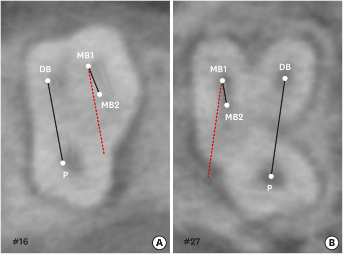
Figure 1 Measurement of the angle between the line connecting the MB1-MB2 and that connecting the DB-P canals in a maxillary molar. (A) Positive angle; (B) Negative angle.MB1, first mesiobuccal root; MB2, second mesiobuccal root; DB, distobuccal; P, palatal.
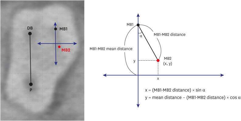
Figure 2 Measurement of the mean distance between the MB1 and MB2 canals in a maxillary molar. The hypothetical line that passes through the MB1 and is parallel to the line connecting the DB and P canals (mm) is the y-axis. The maximum value of the y-axis and the 0 point are assumed to be the mean distance between the MB1 and MB2 canals.MB1, first mesiobuccal root; MB2, second mesiobuccal root; DB, distobuccal; P, palatal.
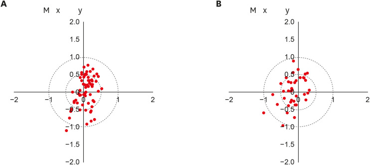
Figure 3 Distribution of the second mesiobuccal root canal in the maxillary first and second molars. (A) Maxillary first molar; (B) Maxillary second molar.
Reference
-
1. Cantatore G, Berutti E, Castellucci A. Missed anatomy: frequency and clinical impact. Endod Topics. 2006; 15:3–31.
Article2. Vertucci FJ. Root canal anatomy of the human permanent teeth. Oral Surg Oral Med Oral Pathol. 1984; 58:589–599. PMID: 6595621.
Article3. Huumonen S, Kvist T, Gröndahl K, Molander A. Diagnostic value of computed tomography in re-treatment of root fillings in maxillary molars. Int Endod J. 2006; 39:827–833. PMID: 16948669.
Article4. Wolcott J, Ishley D, Kennedy W, Johnson S, Minnich S, Meyers J. A 5 yr clinical investigation of second mesiobuccal canals in endodontically treated and retreated maxillary molars. J Endod. 2005; 31:262–264. PMID: 15793380.
Article5. Cotton TP, Geisler TM, Holden DT, Schwartz SA, Schindler WG. Endodontic applications of cone-beam volumetric tomography. J Endod. 2007; 33:1121–1132. PMID: 17931947.6. Michetti J, Maret D, Mallet JP, Diemer F. Validation of cone beam computed tomography as a tool to explore root canal anatomy. J Endod. 2010; 36:1187–1190. PMID: 20630296.
Article7. Blattner TC, George N, Lee CC, Kumar V, Yelton CD. Efficacy of cone-beam computed tomography as a modality to accurately identify the presence of second mesiobuccal canals in maxillary first and second molars: a pilot study. J Endod. 2010; 36:867–870. PMID: 20416435.
Article8. Vizzotto MB, Silveira PF, Arús NA, Montagner F, Gomes BP, da Silveira HE. CBCT for the assessment of second mesiobuccal (MB2) canals in maxillary molar teeth: effect of voxel size and presence of root filling. Int Endod J. 2013; 46:870–876. PMID: 23442087.
Article9. Akbarzadeh N, Aminoshariae A, Khalighinejad N, Palomo JM, Syed A, Kulild JC, Sadeghi G, Mickel A. The association between the anatomic landmarks of the pulp chamber floor and the prevalence of middle mesial canals in mandibular first molars: an in vivo analysis. J Endod. 2017; 43:1797–1801. PMID: 28864218.
Article10. Lofthag-Hansen S, Huumonen S, Gröndahl K, Gröndahl HG. Limited cone-beam CT and intraoral radiography for the diagnosis of periapical pathology. Oral Surg Oral Med Oral Pathol Oral Radiol Endod. 2007; 103:114–119. PMID: 17178504.
Article11. Matherne RP, Angelopoulos C, Kulild JC, Tira D. Use of cone-beam computed tomography to identify root canal systems in vitro . J Endod. 2008; 34:87–89. PMID: 18155501.
Article12. Jang JH, Kim JW, Cho KM, Kim SY, Park SH. A study on Radix Entomolaris about prevalence and correlation of canal orifices location according to number of roots in mandibular first molars. J Korean Dent Assoc. 2018; 56:695–706.13. Deutsch AS, Musikant BL, Gu S, Isidro M. Morphological measurements of anatomic landmarks in pulp chambers of human maxillary furcated bicuspids. J Endod. 2005; 31:570–573. PMID: 16044038.
Article14. Degerness RA, Bowles WR. Dimension, anatomy and morphology of the mesiobuccal root canal system in maxillary molars. J Endod. 2010; 36:985–989. PMID: 20478451.
Article15. Martins JN, Alkhawas MA, Altaki Z, Bellardini G, Berti L, Boveda C, Chaniotis A, Flynn D, Gonzalez JA, Kottoor J, Marques MS, Monroe A, Ounsi HF, Parashos P, Plotino G, Ragnarsson MF, Aguilar RR, Santiago F, Seedat HC, Vargas W, von Zuben M, Zhang Y, Gu Y, Ginjeira A. Worldwide analyses of maxillary first molar second mesiobuccal prevalence: a multicenter cone-beam computed tomographic study. J Endod. 2018; 44:1641–1649.e1. PMID: 30243661.
Article16. Lee YS. Prevalence and location of the second mesiobuccal canal in maxillary first molars in a Korean population. Daegu: Kyungpook National University;2018.17. Betancourt P, Navarro P, Cantín M, Fuentes R. Cone-beam computed tomography study of prevalence and location of MB2 canal in the mesiobuccal root of the maxillary second molar. Int J Clin Exp Med. 2015; 8:9128–9134. PMID: 26309568.18. Lee JH, Kim KD, Lee JK, Park W, Jeong JS, Lee Y, Gu Y, Chang SW, Son WJ, Lee WC, Baek SH, Bae KS, Kum KY. Mesiobuccal root canal anatomy of Korean maxillary first and second molars by cone-beam computed tomography. Oral Surg Oral Med Oral Pathol Oral Radiol Endod. 2011; 111:785–791. PMID: 21439860.
Article19. Bauman R, Scarfe W, Clark S, Morelli J, Scheetz J, Farman A. Ex vivo detection of mesiobuccal canals in maxillary molars using CBCT at four different isotropic voxel dimensions. Int Endod J. 2011; 44:752–758. PMID: 21470249.
Article20. Kim Y, Lee SJ, Woo J. Morphology of maxillary first and second molars analyzed by cone-beam computed tomography in a Korean population: variations in the number of roots and canals and the incidence of fusion. J Endod. 2012; 38:1063–1068. PMID: 22794206.
Article21. Zhang R, Yang H, Yu X, Wang H, Hu T, Dummer PM. Use of CBCT to identify the morphology of maxillary permanent molar teeth in a Chinese subpopulation. Int Endod J. 2011; 44:162–169. PMID: 21091495.
Article22. Kulild JC, Peters DD. Incidence and configuration of canal systems in the mesiobuccal root of maxillary first and second molars. J Endod. 1990; 16:311–317. PMID: 2081944.
Article
- Full Text Links
-
- Actions
-
Cited
- CITED
-
- Close
- Share
-
- Similar articles
-
- Analysis of C-shaped root canal configuration in maxillary molars in a Korean population using cone-beam computed tomography
- Assessment of the relationship between the maxillary molars and adjacent structures using cone beam computed tomography
- A cone-beam computed tomographic study of C-shaped root and root canal in maxillary molars
- Apical periodontitis in mesiobuccal roots of maxillary molars: influence of anatomy and quality of root canal treatment, a CBCT study
- Evaluation of root and root canal morphology of elderly Korean patients maxillary molarsusing cone-beam computed tomography
