Crystal Deposits on Renal Papillae in Stone Formers
- Affiliations
-
- 1Department of Urology, Chonbuk National University Medical School, Jeonju, Korea. ygkim@chonbuk.ac.kr
Abstract
- PURPOSE
Papillary "Randall's plaques" are theorized to act as nidi for urinary stone formation. In this study, we used endoscopic examination and intraoperative biopsy of renal papilla in stone formers undergoing percutaneous nephrolithotomy (PNL) to investigate the correlation between Randall's papillary plaque and primary stone composition and metabolic risk factors.
MATERIALS AND METHODS
A total of 34 patients with renal stones were enrolled. During PNL performed for stone removal, biopsy specimens were taken from selected papilla. We evaluated constituents such as volume, sodium, uric acid, calcium, oxalate, and citrate from 24-hour urine samples, and calcium, sodium, uric acid, phosphate, potassium, and chloride from serum samples 1 month after PNL.
RESULTS
We identified Randall's plaque in 26 patients as irregular, whitish lesions, generally located on the papillary tip. We performed intraoperative biopsies of papilla in the kidneys of stone formers and of known regions of crystal deposits in the interstitial tissue surrounding the ducts. There was no correlation between serum variables, 24-hour urine constituents, and presence of plaque. However, 24-hour urine volume was negatively correlated with the presence of plaque. The incidence of papillary plaques varied with the primary composition of extracted stones and was 80% for calcium oxalate, 92% for calcium phosphate, 50% for uric acid, and 25% for struvite stones (p=0.035).
CONCLUSIONS
The incidence of papillary Randall's plaques in patients with nephrolithiasis varied with the primary composition of formed urinary stones. Randall's plaques are found in most patients with calcium stones. Our findings suggest that the presence of papillary plaque is associated with calcium nephrolithiasis and may contribute to the pathogenesis, treatment, and prevention of calcium urinary stones.
MeSH Terms
-
Biopsy
Calcium
Calcium Oxalate
Calcium Phosphates
Citric Acid
Humans
Incidence
Kidney
Lithotripsy
Magnesium Compounds
Nephrolithiasis
Nephrostomy, Percutaneous
Phosphates
Potassium
Risk Factors
Sodium
Uric Acid
Urinary Calculi
Calcium
Calcium Oxalate
Calcium Phosphates
Citric Acid
Magnesium Compounds
Phosphates
Potassium
Sodium
Uric Acid
Figure
-
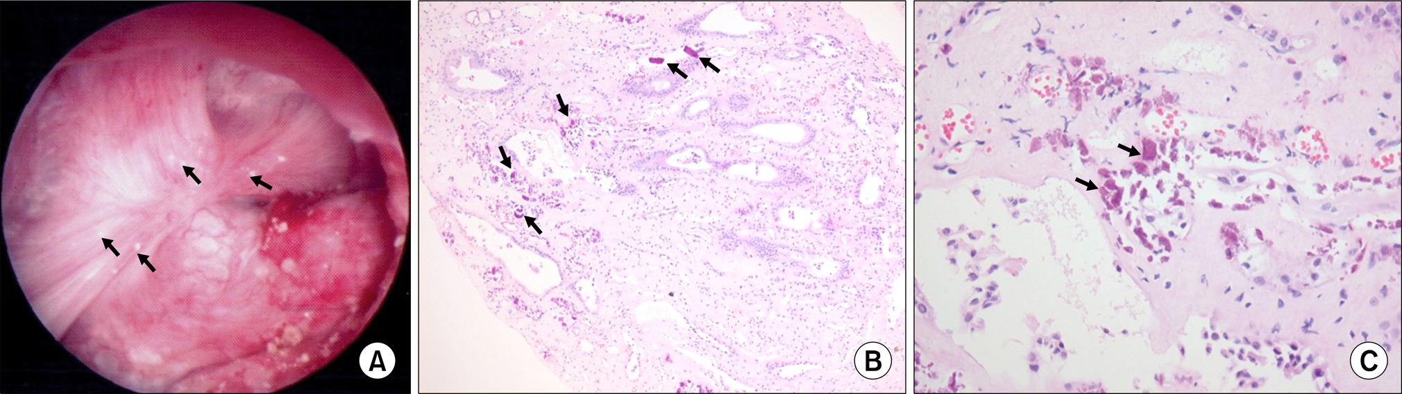
Fig. 1. Endoscopic and histologic images of Randall’s plaques in stone formers. In (A), an example of a papilla from a stone former that was videotaped at the time of the mapping is shown. Several sites of Randall’s plaque (arrows) appear as irregular white areas beneath the urothelium. In addition, a plaque site was noted that lacked a urothelial layer and was thought to be a site where a stone had been attached to the side of the papilla (arrows). In (B), a low-magnification light-microscopic image of a papillary biopsy specimen from a patient is shown; the sites of calcium deposits are indicated with arrows. (C) A light micrograph shows large regions of crystal deposits in the interstitial tissue surrounding the ducts (arrows). H&E stain, magnification, x100 (B); x400 (C).
Reference
-
1.Porena M., Guiggi P., Micheli C. Prevention of stone disease. Urol Int. 2007. 79(Suppl 1):37–46.
Article2.Bartoletti R., Cai T., Mondaini N., Melone F., Travaglini F., Carini M, et al. Epidemiology and risk factors in urolithiasis. Urol Int. 2007. 79(Suppl 1):3–7.
Article3.Pearle MS., Calhoun EA., Curhan GC. Urologic diseases in America project: urolithiasis. J Urol. 2005. 173:848–57.
Article4.Randall A. The origin and growth of renal calculi. Ann Surg. 1937. 105:1009–27.
Article5.Matlaga BR., Williams JC Jr., Kim SC., Kuo RL., Evan AP., Bledsoe SB, et al. Endoscopic evidence of calculus attachment to Randall's plaque. J Urol. 2006. 175:1720–4.
Article6.Low RK., Stoller ML. Endoscopic mapping of renal papillae for Randall's plaques in patients with urinary stone disease. J Urol. 1997. 158:2062–4.
Article7.Indridason OS., Birgisson S., Edvardsson VO., Sigvaldason H., Sigfusson N., Palsson R. Epidemiology of kidney stones in Iceland: a population-based study. Scand J Urol Nephrol. 2006. 40:215–20.
Article8.Ramello A., Vitale C., Marangella M. Epidemiology of nephrolithiasis. J Nephrol. 2000. 13(Suppl 3):S45–50.9.Trinchieri A., Coppi F., Montanari E., Del Nero A., Zanetti G., Pisani E. Increase in the prevalence of symptomatic upper urinary tract stones during the last ten years. Eur Urol. 2000. 37:23–5.
Article10.Ye SJ., Yoo ES., Park YK. Analysis of urinary stone components during the last two decades. Korean J Urol. 2007. 48:1285–8.
Article11.Jun IO., Moon YT. Comparison of stone metabolic risk factors in recurrent stone formers according to sex and age. Korean J Urol. 2002. 43:733–7.12.Kim SD., Yang WJ., Chung JY. Recurrence rate and risk factors for stone recurrence after successful extracorporeal shock wave lithotripsy: 5-year-follow-up study. Korean J Urol. 2007. 48:49–53.
Article13.Abe T., Akakura K., Kawaguchi M., Ueda T., Ichikawa T., Ito H, et al. Outcomes of shockwave lithotripsy for upper urinary-tract stones: a large-scale study at a single institution. J Endourol. 2005. 19:768–73.
Article14.Netelenbos JC., Zwijnenburg PJ., ter Wee PM. Risk factors determining active urinary stone formation in patients with urolithiasis. Clin Nephrol. 2005. 63:188–92.
Article15.DeFoor W., Minevich E., Jackson E., Reddy P., Clark C., Sheldon C, et al. Urinary metabolic evaluations in solitary and recurrent stone forming children. J Urol. 2008. 179:2369–72.
Article16.Robinson MR., Leitao VA., Haleblian GE., Scales CD Jr., Chandrashekar A., Pierre SA, et al. Impact of long-term potassium citrate therapy on urinary profiles and recurrent stone formation. J Urol. 2009. 181:1145–50.
Article17.Cha JS., Jeon SB., Kim MK., Jeong YB., Kim YG. Metabolic stone risk factors associated with papillary calcification on unenhanced spiral computed tomography. Korean J Urol. 2006. 47:507–11.
Article18.Kuo RL., Lingeman JE., Evan AP., Paterson RF., Parks JH., Bledsoe SB, et al. Urine calcium and volume predict coverage of renal papilla by Randall's plaque. Kidney Int. 2003. 64:2150–4.
Article19.Evan AP., Lingeman JE., Coe FL., Parks JH., Bledsoe SB., Shao Y, et al. Randall's plaque or patients with nephrolithiasis begins in basement membranes of thin loops of Henle. J Clin Invest. 2003. 111:607–16.20.Miller NL., Gillen DL., Williams JC Jr., Evan AP., Bledose SB., Coe FL, et al. A formal test of the hypothesis that idiopathic calcium oxalate stones grow on Randall's plaque. BJU Int. 2009. 103:966–71.
Article21.Cifuentes Delatte L., Miñón-Cifuentes J., Medina JA. New studies on papillary calculi. J Urol. 1987. 137:1024–9.22.Low RK., Stoller ML., Schreiber CK. Metabolic and urinary risk factors associated with Randall's papillary plaques. J Endourol. 2000. 14:507–10.
Article
- Full Text Links
-
- Actions
-
Cited
- CITED
-
- Close
- Share
-
- Similar articles
-
- Study on Uric Acid Level in Urinary Calcium Stone Formers
- Study on Serum and Urinary Calcium Level and Serum Parathyroid Hormone in Patients with Urinary Stone
- Study on Serum and Urinary Uric Acid Level in Patients with Urinary Stone
- Comparative study on the morphology of renal pelvoclyceal systems of healthy persons and urinary stone formers by excretory urography
- Comparison of Metabolic Risk Factors in Urolithiasis Patients according to Family History
