The Availability of Radiological Measurement of Femoral Anteversion Angle: Three-Dimensional Computed Tomography Reconstruction
- Affiliations
-
- 1Department of Rehabilitation Medicine, Gyeongsang National University Hospital, Jinju, Korea. virgoperidot@hanmail.net
- KMID: 2309922
- DOI: http://doi.org/10.5535/arm.2016.40.2.237
Abstract
OBJECTIVE
To assess the intra-rater and inter-rater reliability for measuring femoral anteversion angle (FAA) by a radiographic method using three-dimensional computed tomography reconstruction (3D-CT).
METHODS
The study included 82 children who presented with intoeing gait. 3D-CT data taken between 2006 and 2014 were retrospectively reviewed. FAA was measured by 3D-CT. FAA is defined as the angle between the long axis of the femur neck and condylar axis of the distal femur. FAA measurement was performed twice at both lower extremities by each rater. The intra-rater and inter-rater reliability were calculated by intraclass correlation coefficient (ICC).
RESULTS
One hundred and sixty-four lower limbs of 82 children (31 boys and 51 girls, 6.3±3.2 years old) were included. The ICCs of intra-rater measurement for the angle of femoral neck axis (NA) were 0.89 for rater A and 0.96 for rater B, and those of condylar axis (CA) were 0.99 for rater A and 0.99 for rater B, respectively. The ICC of inter-rater measurement for the angle of NA was 0.89 and that of CA was 0.92. By each rater, the ICCs of the intrarater measurement for FAA were 0.97 for rater A and 0.95 for rater B, respectively and the ICC of the inter-rater measurement for FAA was 0.89.
CONCLUSION
The 3D-CT measures for FAA are reliable within individual raters and between different raters. The 3D-CT measures of FAA can be a useful method for accurate diagnosis and follow-up of femoral anteversion.
Keyword
MeSH Terms
Figure
-
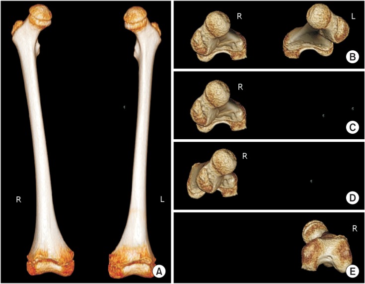
Fig. 1 (A) Anteroposterior view was acquired from 3-dimensional computed tomography. (B) Ninety degrees of rotation by the program resulted in the horizontal view, which is the looking down view of the femur. To measure each FAA, left side femur was eliminated (C) and the remaining right femur was rotated as vertically as possible in a view that hides the femur shaft (D). Upside down view of the femur was rotated to make femur neck as the most linear, for the straightest line (E). R, right; L, left; FAA, femoral anteversion angle.
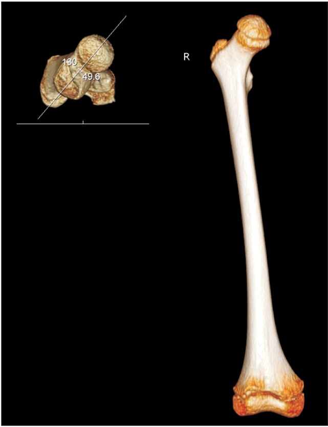
Fig. 2 The angle between the long axis of the femur neck and horizontal line is defined as femoral neck angle. R, right.
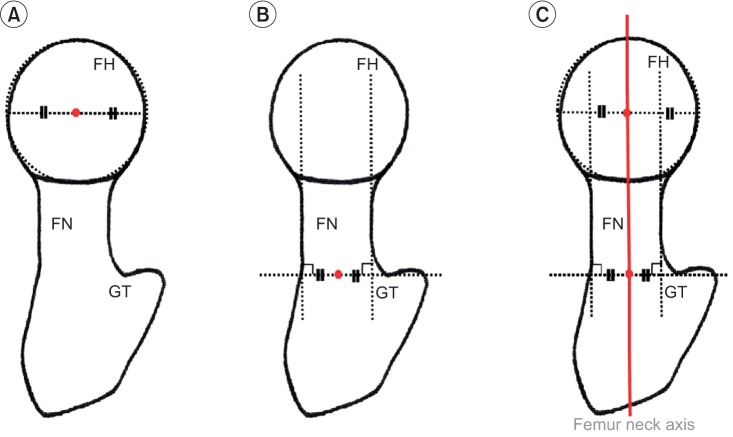
Fig. 3 Definition of femur neck axis. From superior view of 3-dimensional computed tomography, a diagram was constructed. Femur was hypothesized as a circle, and a line that links two points of the circle and passed the center of the circle was drawn (A). Femur neck was hypothesized as a cylinder, and two lines extended from cylinder were drawn. Then, one horizontal line that was closest to the greater trochanter from femur neck and tangential to the two extended lines, was drawn. A point that bisected this line is the center of neck (B). Femur neck axis (thick red line) was defined as a line that connected the two central points (C). FH, femur head; FN, femur neck; GT, greater trochanter.
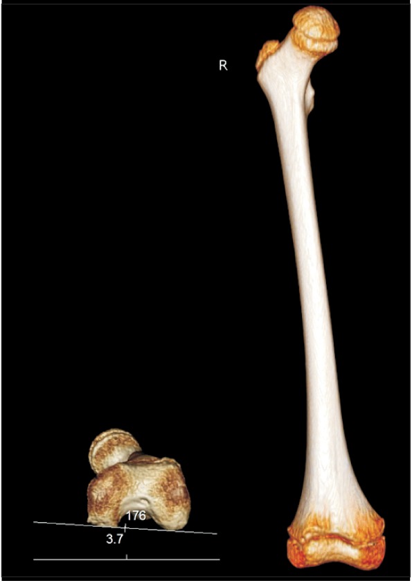
Fig. 4 The angle between the intercondylar axis of the distal femur and horizontal line is defined as condylar angle. R, right.
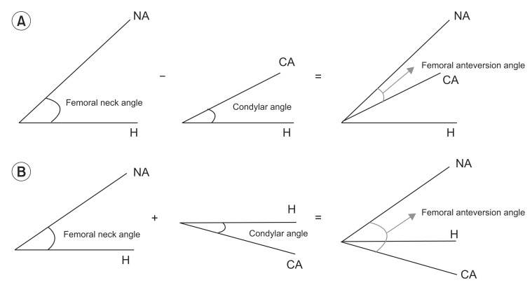
Fig. 5 Femoral anteversion angle was calculated as subtraction of the femoral neck and condylar angles when the two horizontal axis were in the same direction (A), and as summation when the two angles were in the opposite direction (B). H, horizontal axis; NA, femoral neck axis; CA, condylar axis.
Cited by 2 articles
-
Change of Femoral Anteversion Angle in Children With Intoeing Gait Measured by Three-Dimensional Computed Tomography Reconstruction: One-Year Follow-Up Study
Minsik Kong, Hongsik Jo, Chang Han Lee, Se-Woong Chun, Chulho Yoon, Heesuk Shin
Ann Rehabil Med. 2018;42(1):137-144. doi: 10.5535/arm.2018.42.1.137.Change of Femoral Anteversion Angle in Children With Intoeing Gait Measured by Three-Dimensional Computed Tomography Reconstruction: 3-Year Follow-Up Study
Yeongchae Park, Hayoung Byun, Mi-Ji Kim, Heesuk Shin
Ann Rehabil Med. 2023;47(3):182-191. doi: 10.5535/arm.23043.
Reference
-
1. Kling TF Jr, Hensinger RN. Angular and torsional deformities of the lower limbs in children. Clin Orthop Relat Res. 1983; 176:136–147. PMID: 6851317.
Article2. Kumar SJ, MacEwen GD. Torsional abnormalities in children's lower extremities. Orthop Clin North Am. 1982; 13:629–639. PMID: 7099592.
Article3. Cibulka MT. Determination and significance of femoral neck anteversion. Phys Ther. 2004; 84:550–558. PMID: 15161420.
Article4. Bobroff ED, Chambers HG, Sartoris DJ, Wyatt MP, Sutherland DH. Femoral anteversion and neck-shaft angle in children with cerebral palsy. Clin Orthop Relat Res. 1999; 364:194–204. PMID: 10416409.
Article5. Gelberman RH, Cohen MS, Desai SS, Griffin PP, Salamon PB, O'Brien TM. Femoral anteversion. A clinical assessment of idiopathic intoeing gait in children. J Bone Joint Surg Br. 1987; 69:75–79. PMID: 3818738.
Article6. Lee DY, Lee CK, Cho TJ. A new method for measurement of femoral anteversion: a comparative study with other radiographic methods. Int Orthop. 1992; 16:277–281. PMID: 1428343.7. Anda S, Terjesen T, Sundalsfoll S, Tangerud A. Femoral anteversion measured by ultrasonography and radiography: an anatomic investigation. Acta Radiol. 1988; 29:695–699. PMID: 3056474.8. Guenther KP, Tomczak R, Kessler S, Pfeiffer T, Puhl W. Measurement of femoral anteversion by magnetic resonance imaging: evaluation of a new technique in children and adolescents. Eur J Radiol. 1995; 21:47–52. PMID: 8654459.9. Lee S, Choi KS, Jeung IS, Lee JE, Yang SM, Lee SM. Physical examination and computed tomography in children with toe in gait. J Korean Acad Rehabil Med. 2011; 35:61–66.10. Kim HD, Lee DS, Eom MJ, Hwang JS, Han NM, Jo GY. Relationship between physical examinations and two-dimensional computed tomographic findings in children with intoeing gait. Ann Rehabil Med. 2011; 35:491–498. PMID: 22506164.
Article11. Weiner DS, Cook AJ, Hoyt WA Jr, Oravec CE. Computed tomography in the measurement of femoral anteversion. Orthopedics. 1978; 1:299–306. PMID: 733194.
Article12. Miller F, Liang Y, Merlo M, Harcke HT. Measuring anteversion and femoral neck-shaft angle in cerebral palsy. Dev Med Child Neurol. 1997; 39:113–118. PMID: 9062426.
Article13. Gose S, Sakai T, Shibata T, Murase T, Yoshikawa H, Sugamoto K. Morphometric analysis of the femur in cerebral palsy: 3-dimensional CT study. J Pediatr Orthop. 2010; 30:568–574. PMID: 20733422.14. Kim SI, Park SB, Lee KM. Measurement of femoral anteversion using 3 dimensional imaging. J Korean Acad Rehabil Med. 1994; 18:495–499.15. Flohr TG, Schaller S, Stierstorfer K, Bruder H, Ohnesorge BM, Schoepf UJ. Multi-detector row CT systems and image-reconstruction techniques. Radiology. 2005; 235:756–773. PMID: 15833981.
Article16. Rengier F, Mehndiratta A, von Tengg-Kobligk H, Zechmann CM, Unterhinninghofen R, Kauczor HU, et al. 3D printing based on imaging data: review of medical applications. Int J Comput Assist Radiol Surg. 2010; 5:335–341. PMID: 20467825.
Article17. Kim JS, Park TS, Park SB, Kim JS, Kim IY, Kim SI. Measurement of femoral neck anteversion in 3D. Part 1: 3D imaging method. Med Biol Eng Comput. 2000; 38:603–609. PMID: 11217876.
Article18. Flohr TG, Schaller S, Stierstorfer K, Bruder H, Ohnesorge BM, Schoepf UJ. Multi-detector row CT systems and image-reconstruction techniques. Radiology. 2005; 235:756–773. PMID: 15833981.
Article
- Full Text Links
-
- Actions
-
Cited
- CITED
-
- Close
- Share
-
- Similar articles
-
- Relationship between Femoral Anteversion and Tibial Torsion in Intoeing Gait
- Measurement of Femoral Anteversion in the PACS Image Viewer
- Comparative Study of the Roentgenographic Methods for the Measurement of the Femoral Anteversion
- Computed Tomography in Evaluation of Femoral Anteversion: Transverse Section Versus Axial Oblique Section
- Change of Femoral Anteversion during Closed Femoral Intramedullary Nailing
