The Efficiency of Laparoscopic Splenorenal Shunt: a Chronic Canine Model
- Affiliations
-
- 1Department of Urology, Dong-A University School of Medicine, Busan, Korea. sunggt@daunet.donga.ac.kr
- KMID: 2294181
- DOI: http://doi.org/10.4111/kju.2006.47.3.316
Abstract
-
PURPOSE: Splenorenal bypass is a major surgical procedure that's used for the management of renal artery stenosis. Herein, we evaluate the feasibility and efficacy of performing laparoscopic splenorenal bypass in a chronic canine model.
MATERIALS AND METHODS
A total of 12 animals were used for this study. The initial 6 acute animals were used to develop the technique. The remaining 6 surviving animals, which form the basis for this report, were used for a chronic study with up to 2 months follow-up. The renal artery and the distal splenic artery was dissected, its proximal end clamped and its distal end cut and spatulated. An end-to-end anastomosis of the splenic artery and renal artery was performed using only laparoscopic freehand suturing and knot-tying techniques. Upon revascularization, a laparoscopic doppler ultrasound probe was used to document blood flow in the renal artery. Three animals were each followed for 1 month and 2 months, respectively.
RESULTS
The total operative time was 297+/-36 min. The mean number of suture bites per anastomosis was 14.3. The only intraoperative complication was hemorrhage from the anastomotic site. Intraoperative Doppler ultrasound documented good blood flow in all 6 animals upon releasing the clamp. At the time of euthanasia, intravenous pyelography (IVP) showed early visualization of the left kidney with prompt drainage in 5 of the 6 surviving animals. In one animal that had two left renal arteries, a distal thrombosis was found despite the patent anastomotic site.
CONCLUSIONS
Laparoscopic splenorenal bypass can be performed in a reproducible fashion with using only intracorporeal techniques. We believe that with experience, complex urologic vascular procedures can be laparoscopically performed in the future.
Keyword
MeSH Terms
Figure
-
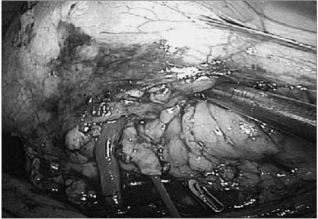
Fig. 1 Intracorporeal renal hypothermia. A 3Fr arterial balloon catheter is inserted through the left renal artery. Intracorporeal renal hypothermia is achieved in situ via an intra-arterial perfusion of ice-cold saline solution through the catheter.
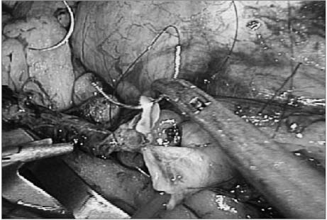
Fig. 2 End-to-end anastomosis between the mobilized splenic artery and the left renal artery with using 6-0 Prolene suture (RB-2 needle) is performed with freehand suturing and knot-tying techniques. During the performance of the anterior wall anastomosis, the arterial catheter is maintained intact within the renal artery, thus maintaining the kidney in a cold ischemia condition.
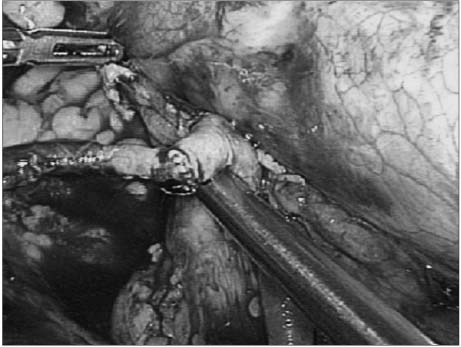
Fig. 3 Revascularization of the kidney. Anastomosis is done in a water-tight manner. The renal arterial blood flow is confirmed by placing a 5mm laparoscopic color Doppler ultrasound probe at the renal artery distal to the anastomotic site.
Reference
-
1. Andrew CN, Amr F. Novick AC, editor. Renovascular hypertension and ischemic nephropathy. Arm Fergany Campbell's urology. 2002. 8th ed. Philadelphia: Saunders;229–271.2. Clayman RV, Kavoussi LR, Soper NJ, Dierks SM, Meretyk S, Darcy MD, et al. Laparoscopic nephrectomy: initial case report. J Urol. 1991. 146:278–282.3. Kim HH. Laparoscopic surgery in urology. Korean J Urol. 2003. 44:945–948.4. Baldwin DD, Maynes LJ, Berger KA, Desai PJ, Zuppan CW, Zimmerman GJ, et al. Laparoscopic warm renal ischemia in the solitary porcine kidney model. Urology. 2004. 64:592–597.5. Lim DJ, Kim HH, Moon YT, Park YY, Yang SK, Yoon SJ, et al. Endourologic procedures and laparoscopic surgery in urology training hospitals: the report of nationwide survey. Korean J Urol. 2004. 45:714–719.6. Kong GS, Seong YK, Sung GT. Robotic-assisted radical prostatectomy using da Vinci™ surgical robotic system: initial Korean experience. Korean J Urol. 2005. 46:353–359.7. Seo IY, Park SC, Oh SJ. Laparoscopic pyeloplasty with transperitoneal approach for ureteropelvic obstruction. Korean J Urol. 2005. 46:370–374.8. Heo J, Kim TH, Kong GS, Sung GT. The initial experience of laparoscopic ureteroureterostomy for retrocaval ureter. Korean J Urol. 2005. 46:422–425.9. Kim HH, Kim GH, Sung GT. Laparoscopic adrenalectomy for pheochromocytoma: comparison with conventional open adrenalectomy. J Endourol. 2004. 18:251–255.10. Abelson DS, Haimovici H, Hurwitt ES, Seidenberg B. Splenorenal arterial anastomoses. Circulation. 1956. 14:532–539.11. Khauli RB, Novick AC, Ziegelbaum M. Splenorenal bypass in the treatment of renal artery stenosis: experience with sixty-nine cases. J Vasc Surg. 1985. 2:547–551.12. Moncure AC, Brewster DC, Darling RC, Atnip RG, Newton WD, Abbott WM. Use of the splenic and hepatic arteries for renal revascularization. J Vasc Surg. 1986. 3:196–203.13. Geroulakos G, Wright JG, Tober JC, Anderson L, Smead WL. Use of the splenic and hepatic artery for renal revascularization in patients with atherosclerotic renal artery disease. Ann Vasc Surg. 1997. 11:85–89.14. Hsu TH, Gill IS, Sung GT, Meraney A, McMahon JT, Novick AC. Laparoscopic aortorenal bypass. J Endourol. 2000. 14:123–131.15. Picod G, Jambon AC, Vinatier D, Dubois P. What can the operator actually feel when performing a laparoscopy? Surg Endosc. 2005. 19:95–100.16. Rosenberg BH, Landsittel D, Averch TD. Can video games be used to predict or improve laparoscopic skills? J Endourol. 2005. 19:372–376.17. Fergany AF, Gill IS, Kaouk JH, Meraney AM, Hafez KS, Sung GT. Laparoscopic intracorporeally constructed ileal conduit after porcine cystoprostatectomy. J Urol. 2001. 166:285–288.
- Full Text Links
-
- Actions
-
Cited
- CITED
-
- Close
- Share
-
- Similar articles
-
- A case of post-operative chylous ascites after a splenorenal shunt operation in a child with congenital hepatic fibrosis
- Epidural Analgesia in a Parturient following Splenorenal Shunt Operation for Liver Cirrhosis
- Distal pancreatectomy with splenorenal shunt to preserve spleen in a cirrhotic patient
- Lessons Learned from Inappropriate Ligation of the Left Renal Vein for a Large Splenorenal Shunt in Living Donor Liver Transplantation
- Living-Donor Liver Transplantation with Renoportal Anastomosis using an Interposition Polytetrafluoroethylene Graft for a Patient with Large Spontaneous Splenorenal Shunt: A Case Report
