J Breast Cancer.
2010 Mar;13(1):83-89. 10.4048/jbc.2010.13.1.83.
Clinicopathological Characteristics in Invasive Ductal Breast Cancer with Low FDG Uptake in (18)F-FDG PET/CT
- Affiliations
-
- 1Department of Surgery, Pusan National University School of Medicine, Busan, Korea. bytae@pusan.ac.kr
- 2Department of Nuclear Medicine, Pusan National University School of Medicine, Busan, Korea.
- 3Department of Pathology, Pusan National University School of Medicine, Busan, Korea.
- KMID: 2286564
- DOI: http://doi.org/10.4048/jbc.2010.13.1.83
Abstract
- PURPOSE
The aim of this study is to evaluate whether low FDG uptake would be associated with the biological low-aggressiveness of invasive ductal carcinoma.
METHODS
The subjects consisted of 124 female patients with primary invasive ductal carcinoma. All the patients were examined with (18)F-FDG PET/CT before neoadjuvant chemotherapy.
RESULTS
With regard to histopathologic grading, 117 were histopathologic grade 1 and 2, and 7 were grade 3. Low FDG uptake correlated with well and moderate histopathologic grade (p=0.003) and low (18)F-FDG uptake in invasive ductal carcinoma depended on the presence of axillary lymph node metastases (p=0.014) and small tumor (<2.0 cm, p=0.022). Ki-67 positivity ranged from 0% to 60% (mean 15%). Sixty seven specimens showed low immunoreactivity to Ki-67 antigen (<10% of tumor cells). This revealed a significant correlation between low FDG uptake and Ki-67 (p=0.003). Logistic regression analysis between these factors showed that lower histologic grade, no axillary lymph nodes metastases and low Ki-67 (<10%) were correlated with low FDG uptake.
CONCLUSION
Our results demonstrated that an association exists between low FDG uptake and good prognostic factors such as lower histologic grade (1, 2), no axillary lymph node metastases and low Ki-67 (<10%).
MeSH Terms
Figure
-
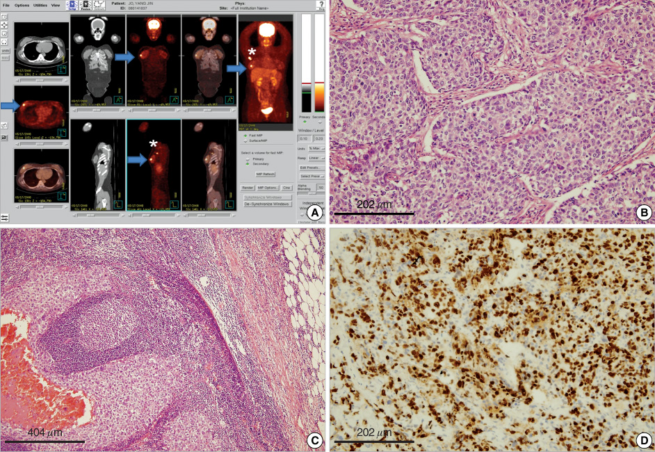
Figure 1 (A) 18FDG PET/CT shows increased FDG uptake of right breast (arrow) and ipsilateral axillary LN (*). (B) Microscopic appearance of the specimen shows grade 3. (C) It is metastasized to axillary lymph node. (D) Ki-67 ratio is 40%.
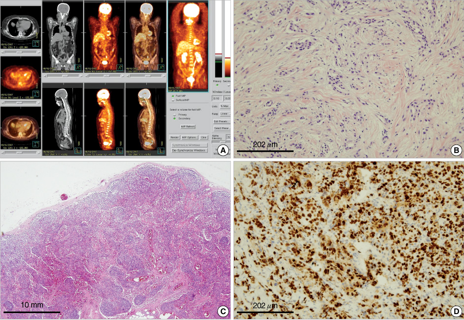
Figure 2 (A) It is a false-negative finding at 18F-FDG-PET/CT during staging of left breast cancer in 58-yr-old woman and 18F-FDG-PET/CT image shows low FDG uptake. (B) Microscopic appearance of the specimen is grade 1. (C) It doesn't show axillary lymph node metastasis. (D) Ki-67 ratio is 1%.
Reference
-
1. Tse NY, Hoh CK, Hawkins RA, Zinner MJ, Dahlbom M, Choi Y, et al. The application of positron emission tomographic imaging with fluorodeoxyglucose to the evaluation of breast disease. Ann Surg. 1992. 216:27–34.
Article2. Bruce DM, Evans NT, Heys SD, Needham G, BenYounes H, Mikecz P, et al. Positron emission tomography: 2-deoxy-2-[18F]-fluoro-D-glucose uptake in locally advanced breast cancers. Eur J Surg Oncol. 1995. 21:280–283.
Article3. Crippa F, Agresti R, Seregni E, Greco M, Pascali C, Bogni A, et al. Prospective evaluation of (18F)FDG positron emission tomography in the presurgical staging of the axilla in breast cancer. J Nucl Med. 1998. 39:4–8.4. Schirrmeister H, Kühn T, Guhlmann A, Santjohanser C, Hörster T, Nüssle K, et al. Fluorine-18 2-deoxy-2-fluoro-D-glucose PET in the preoperative staging of breast cancer: comparision with the standard staging procedures. Eur J Nucl Med. 2001. 28:351–358.
Article5. Avril N, Dose J, Jänicke F, Bense S, Ziegler S, Laubenbacher C, et al. Metabolic characterization of breast tumors with position emission tomography using F-18 fluorodeoxyglucose. J Clin Oncol. 1996. 14:1848–1857.
Article6. Schomburg A, Bender H, Reichet C, Sommer T, Ruhlmann J, Kozak B, et al. Standardized uptake values of fluorine-18 fluorodeoxyglucose: the value of different normalization procedures. Eur J Nucl Med Mol Imaging. 1996. 23:571–574.
Article7. Adler LP, Crowe JP, al-Kaisi NK, Sunshine JL. Evaluation of breast masses and axillary lymph nodes with [F-18]2-deoxy-2-fluoro-D-glucose PET. Radiology. 1993. 187:743–750.
Article8. Walter C, Scheidhauer K, Scharl A, Goering UJ, Theissen P, Kugel H, et al. Clinical and diagnostic value of preoperative MR mammography and FDG-PET in suspicious breast lesions. Eur Radiol. 2003. 13:1651–1656.
Article9. Rosé C, Dose J, Avril N. Positron emission tomography for the diagnosis of breast cancer. Nucl Med Commun. 2002. 23:613–618.
Article10. Shimoda W, Hayashi M, Murakami K, Oyama T, Sunagawa M. The relationship between FDG uptake in PET scans and biological behavior in breast cancer. Breast Cancer. 2007. 14:260–268.
Article11. Buck A, Schirrmeister H, Kühn T, Shen C, Kalker T, Kotzerke J, et al. FDG uptake in breast cancer: correlation with biological and clinical prognostic parameters. Eur J Nucl Med Mol Imaging. 2002. 29:1317–1323.
Article12. Avril N, Menzel M, Dose J, Schelling M, Weber W, Jänicke F, et al. Glucose metabolism of breast cancer associated by 18F-FDG PET: histologic and immunohistochemical tissue analysis. J Nucl Med. 2001. 42:9–16.13. Weir L, Worsley D, Bernstein V. The value of FDG positron emission tomography in the management of patients with breast cancer. Breast J. 2005. 11:204–209.
Article14. Eubank WB, Mankoff DA. Evolving role of positron emission tomography in breast cancer imaging. Semin Nucl Med. 2005. 35:84–99.
Article15. Lind P, Igerc I, Beyer T, Reinprecht P, Hausegger K. Advantages and limitations of FDG PET in the follow-up of breast cancer. Eur J Nucl Med Mol imaging. 2004. 31:Suppl 1. S125–S134.16. Czernin J. FDG-PET in breast cancer: a different view of its clinical usefulness. Mol Imaging Biol. 2002. 4:35–45.
Article17. Groheux D, Moretti JL, Basillet G, Espie M, Giacchetti S, Hindie E, et al. Effects of (18)F-FDG PET/CT imaging in patients with clinical stage II and III breast cancer. Int J Radiat Oncol Biol Phys. 2008. 71:695–704.
Article18. Gil-Rendo A, Martinez-Regueira F, Zornoza G, Garcia-Velloso MJ, Beorlegui C, Rodriguez-Spiteri N. Association between [18F] fluorodeoxyglucose uptake and prognostic parameters in breast cancer. Br J Surg. 2009. 96:166–170.
Article19. Avril N, Rosé CA, Schelling M, Dose J, Kuhn W, Bense S, et al. Breast imaging with positron emission tomography and fluorine-18 fluorodeoxyglucose: use and limitations. J Clin Oncol. 2000. 18:3495–3502.
Article20. Flope AL, Lyles RH, Sprouse JT, Conrad EU 3rd, Eary JF. (F-18) fluorodeoxyglucose positron emission tomography as a predictor of pathologic grade and other prognostic variables in bone and soft tissue sarcoma. Clin Cancer Res. 2000. 6:1279–1287.21. Crippa F, Seregni E, Agresti R, Chiesa C, Pascali C, Bogni A, et al. Association between [18F]fluorodeoxyglucose uptake and postoperative histopathology, hormone receptor status, thymidine labelling index and p53 in primary breast cancer: a preliminary observation. Eur J Nucl Med. 1998. 25:1429–1434.
Article22. Fisher B, Slack NH. Number of lymph nodes examined and the prognosis of breast carcinoma. Surg Gynecol Obstet. 1970. 131:79–88.23. Ueda S, Tsuda H, Asakawa H, Shigekawa T, Fukatsu K, Kondo N, et al. Clinicopathological and prognostic relevance of uptake level using 18F-fluorodeoxyglucose positron emission tomography/computed tomography fusion imaging (18F-FDG-PET/CT) in primary breast cancer. Jpn J Clin Oncol. 2008. 38:250–258.
Article24. Liberman L. Pathologic analysis of sentinel lymph nodes in breast carcinoma. Cancer. 2000. 88:971–977.
Article25. Bos R, van Der Hoeven JJ, van Der Wall E, van Der Groep P, van Diest P, Comans EF, et al. Biologic correlates of (18)fluorodeoxyglucose uptake in human breast cancer measured by positron emission tomography. J Clin Oncol. 2002. 20:379–387.
Article26. Buck AK, Schirrmeister H, Mattfeldt T, Reske SN. Biological characterisation of breast cancer by means of PET. Eur J Nucl Med Mol Imaging. 2004. 31:Suppl 1. S80–S87.
Article27. Eberlein TJ. Current management of carcinoma of the breast. Ann Surg. 1994. 220:121–136.
Article28. Dehdashti F, Mortimer JE, Siegel BA, Griffeth LK, Bonasera TJ, Fusselman MJ, et al. Positron tomographic assessment of estrogen receptors in breast cancer: a comparison with FDG-PET and in vitro receptor assays. J Nucl Med. 1995. 36:1766–1774.29. Dettmar P, Harbeck N, Thomssen C, Pache L, Ziffer P, Fizi K, et al. Prognosis impact of proliferation-associated factors MIB1 (Ki-67) and S-phase in node-negative breast cancer. Br J Cancer. 1997. 75:1525–1533.
Article
- Full Text Links
-
- Actions
-
Cited
- CITED
-
- Close
- Share
-
- Similar articles
-
- Correlation of Primary Tumor FDG Uptake with Clinicopathologic Prognostic Factors in Invasive Ductal Carcinoma of the Breast
- Uterine Epithelioid Angiosarcoma on F-18 FDG PET/CT
- F-18 FDG PET/CT Finding in Solid Pseudo-papillary Tumor of the Pancreas 6 years After Initial Diagnosis
- (18)F-FDG PET/CT with Contrast Enhancement for Evaluation of Axillary Lymph Node Involvement in T1 Breast Cancer
- Prognostic Value of Primary Tumor Uptake on F-18 FDG PET/CT in Patients with Invasive Ductal Breast Cancer
