Expression and Activity of the Na-K ATPase in Ischemic Injury of Primary Cultured Astrocytes
- Affiliations
-
- 1Department of Physiology, Biomedical Science Institute and Medical Research Center, School of Medicine, Kyung Hee University, Seoul 130-701, Korea. ywcho@khu.ac.kr
- 2Department of Pharmacology, Biomedical Science Institute and Medical Research Center, School of Medicine, Kyung Hee University, Seoul 130-701, Korea.
- KMID: 2285461
- DOI: http://doi.org/10.4196/kjpp.2013.17.4.275
Abstract
- Astrocytes are reported to have critical functions in ischemic brain injury including protective effects against ischemia-induced neuronal dysfunction. Na-K ATPase maintains ionic gradients in astrocytes and is suggested as an indicator of ischemic injury in glial cells. Here, we examined the role of the Na-K ATPase in the pathologic process of ischemic injury of primary cultured astrocytes. Chemical ischemia was induced by sodium azide and glucose deprivation. Lactate dehydrogenase assays showed that the cytotoxic effect of chemical ischemia on astrocytes began to appear at 2 h of ischemia. The expression of Na-K ATPase alpha1 subunit protein was increased at 2 h of chemical ischemia and was decreased at 6 h of ischemia, whereas the expression of alpha1 subunit mRNA was not changed by chemical ischemia. Na-K ATPase activity was time-dependently decreased at 1, 3, and 6 h of chemical ischemia, whereas the enzyme activity was temporarily recovered to the control value at 2 h of chemical ischemia. Cytotoxicity at 2 h of chemical ischemia was significantly blocked by reoxygenation for 24 h following ischemia. Reoxygenation following chemical ischemia for 1 h significantly increased the activity of the Na-K ATPase, while reoxygenation following ischemia for 2 h slightly decreased the enzyme activity. These results suggest that the critical time for ischemia-induced cytotoxicity of astrocytes might be 2 h after the initiation of ischemic insult and that the increase in the expression and activity of the Na-K ATPase might play a protective role during ischemic injury of astrocytes.
MeSH Terms
Figure
-
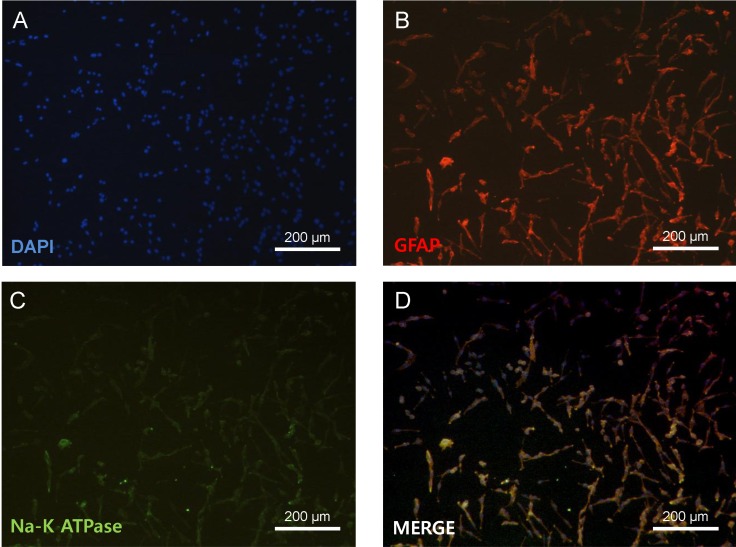
Fig. 1 Primary cultures of astrocytes. Immunohistochemistry of primary astrocytes and Na-K ATPase. DAPI, blue (A); GFAP, red (B); Na-K ATPase, green (C); Merged image (D). Scale bar=200 µm.
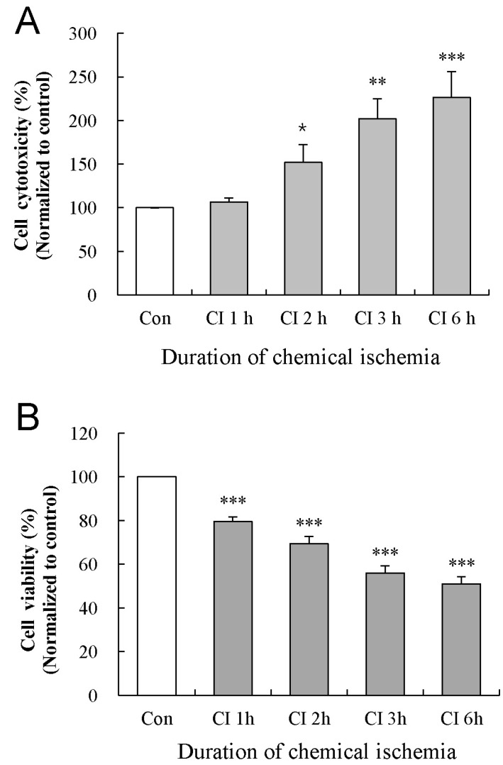
Fig. 2 Effect of chemical ischemia on the cytotoxicity and viability of cultured astrocytes. Cytotoxicity and cell viability was measured by LDH and MTT assays, respectively. The levels of LDH release (A) and MTT reduction (B) were quantified and compared to the control at each time point. Each value indicates the mean±S.E.M. normalized to the control of each time point. Data were obtained from five experiments. *p<0.05, **p<0.01, and ***p<0.001.
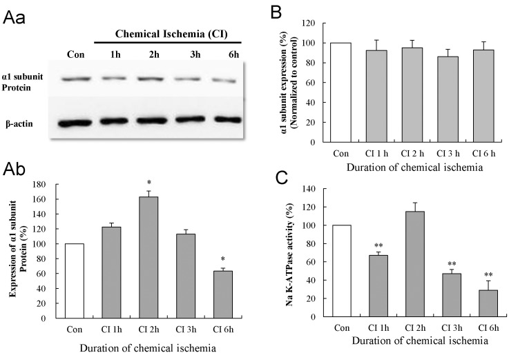
Fig. 3 Effect of chemical ischemia on the expression of Na-K ATPase α1 subunit and Na-K ATPase activity in cultured astrocytes. (A) Western blot analysis shows the expression of Na-K ATPase α1 subunit protein following chemical ischemia for 1, 2, 3, and 6 h. Data were obtained from five experiments. (B) Real-time PCR analysis shows the expression of Na-K ATPase α1 subunit mRNA in cultured astrocytes following chemical ischemia for 1, 2, 3, and 6 h. Data were obtained from four experiments. (C) The activity of Na-K ATPase in the membrane fraction of cultured astrocytes after chemical ischemia for 1, 2, 3, and 6 h. Data were obtained from five experiments. For all experiments, each value indicates the mean±S.E.M. normalized to the control of each time point. *p<0.05, **p<0.01.
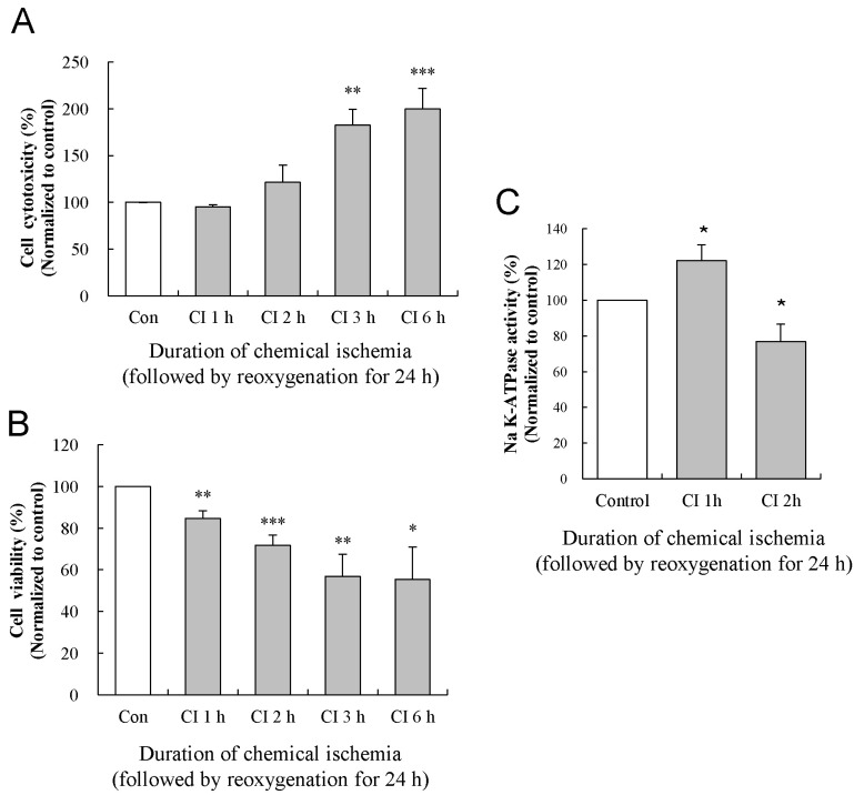
Fig. 4 Effects of reoxygenation on the changes in the cytotoxicity, viability, and activity of the Na-K ATPase. The levels of LDH release (A) and MTT reduction (B) were quantified at 24 h of reoxygenation following chemical ischemia for 1, 2, 3, and 6 h. (C) Activity of Na-K ATPase was measured at 24 h of reoxygenation following chemical ischemia for 1 and 2 h. Data show the mean± S.E.M. of the relative values obtained from five animals. *p<0.05, **p<0.01, and ***p<0.001.
Reference
-
1. Di Carlo A. Human and economic burden of stroke. Age Ageing. 2009; 38:4–5. PMID: 19141505.
Article2. Dugan LL, Kim-Han JS. Astrocyte mitochondria in in vitro models of ischemia. J Bioenerg Biomembr. 2004; 36:317–321. PMID: 15377865.
Article3. Rossi DJ, Brady JD, Mohr C. Astrocyte metabolism and signaling during brain ischemia. Nat Neurosci. 2007; 10:1377–1386. PMID: 17965658.
Article4. Skou JC. The Na,K-pump. Methods Enzymol. 1988; 156:1–25. PMID: 2835594.5. Golden WC, Brambrink AM, Traystman RJ, Martin LJ. Failure to sustain recovery of Na,K-ATPase function is a possible mechanism for striatal neurodegeneration in hypoxic-ischemic newborn piglets. Brain Res Mol Brain Res. 2001; 88:94–102. PMID: 11295235.
Article6. Hernández-R J. Na+/K+-ATPase regulation by neurotransmitters. Neurochem Int. 1992; 20:1–10. PMID: 1363908.7. Specht SC. Development and regional distribution of two molecular forms of the catalytic subunit of the Na+, K+-ATPase in rat brain. Biochem Biophys Res Commun. 1984; 121:208–212. PMID: 6329195.8. Kasai K, Yamashita T, Yamaguchi A, Yoshiya K, Kawakita A, Tanaka H, Sugimoto H, Tohyama M. Induction of mRNAs and proteins for Na/K ATPase alpha1 and beta1 subunits following hypoxia/reoxygenation in astrocytes. Brain Res Mol Brain Res. 2003; 110:38–44. PMID: 12573531.9. Watts AG, Sanchez-Watts G, Emanuel JR, Levenson R. Cell-specific expression of mRNAs encoding Na+, K+-ATPase alpha-and beta-subunit isoforms within the rat central nervous system. Proc Natl Acad Sci U S A. 1991; 88:7425–7429. PMID: 1651505.10. Bezzi P, Volterra A. A neuron-glia signalling network in the active brain. Curr Opin Neurobiol. 2001; 11:387–394. PMID: 11399439.
Article11. Benarroch EE. Neuron-astrocyte interactions: partnership for normal function and disease in the central nervous system. Mayo Clin Proc. 2005; 80:1326–1338. PMID: 16212146.
Article12. Swanson RA, Ying W, Kauppinen TM. Astrocyte influences on ischemic neuronal death. Curr Mol Med. 2004; 4:193–205. PMID: 15032713.
Article13. Stanimirovic DB, Ball R, Durkin JP. Stimulation of glutamate uptake and Na,K-ATPase activity in rat astrocytes exposed to ischemia-like insults. Glia. 1997; 19:123–134. PMID: 9034829.14. Swanson RA, Benington JH. Astrocyte glucose metabolism under normal and pathological conditions in vitro. Dev Neurosci. 1996; 18:515–521. PMID: 8940626.
Article15. Silver IA, Erecińska M. Energetic demands of the Na+/K+ ATPase in mammalian astrocytes. Glia. 1997; 21:35–45. PMID: 9298845.16. Xie M, Wang W, Kimelberg HK, Zhou M. Oxygen and glucose deprivation-induced changes in astrocyte membrane potential and their underlying mechanisms in acute rat hippocampal slices. J Cereb Blood Flow Metab. 2008; 28:456–467. PMID: 17713462.
Article17. Kwon HJ, Hwang IK, An HJ, Han SH, Yang JI, Shin HS, Yoo ID, Kang TC. Changes of glial Na+/K+ ATPase (alpha 1 subunit) immunoreactivity in the gerbil hippocampus after transient forebrain ischemia. Brain Res. 2003; 987:233–239. PMID: 14499968.18. McCarthy KD, de Vellis J. Preparation of separate astroglial and oligodendroglial cell cultures from rat cerebral tissue. J Cell Biol. 1980; 85:890–902. PMID: 6248568.
Article19. Selvatici R, Previati M, Marino S, Marani L, Falzarano S, Lanzoni I, Siniscalchi A. Sodium azide induced neuronal damage in vitro: evidence for non-apoptotic cell death. Neurochem Res. 2009; 34:909–916. PMID: 18841470.20. Bhuiyan MI, Kim HB, Kim SY, Cho KO. The Neuroprotective Potential of Cyanidin-3-glucoside Fraction Extracted from Mulberry Following Oxygen-glucose Deprivation. Korean J Physiol Pharmacol. 2011; 15:353–361. PMID: 22359473.
Article21. Hur J, Lee P, Kim MJ, Kim Y, Cho YW. Ischemia-activated microglia induces neuronal injury via activation of gp91phox NADPH oxidase. Biochem Biophys Res Commun. 2010; 391:1526–1530. PMID: 20036216.
Article22. Scherzer P, Popovtzer MM. Segmental localization of mRNAs encoding Na+/K+-ATPase alpha(1)-and beta(1)-subunits in diabetic rat kidneys using RT-PCR. Am J Physiol Renal Physiol. 2002; 282:F492–F500. PMID: 11832431.23. Mitsumoto Y, Klip A. Development regulation of the subcellular distribution and glycosylation of GLUT1 and GLUT4 glucose transporters during myogenesis of L6 muscle cells. J Biol Chem. 1992; 267:4957–4962. PMID: 1311324.
Article24. Esmann M. ATPase and phosphatase activity of Na+/K+-ATPase: molar and specific activity, protein determination. Methods Enzymol. 1988; 156:105–115. PMID: 2835596.25. Molinas SM, Trumper L, Serra E, Elías MM. Evolution of renal function and Na+/K+-ATPase expression during ischaemia-reperfusion injury in rat kidney. Mol Cell Biochem. 2006; 287:33–42. PMID: 16708288.
- Full Text Links
-
- Actions
-
Cited
- CITED
-
- Close
- Share
-
- Similar articles
-
- Heat Treatment Suppressed the Expression and Activity of Matrix Metalloproteinases in the Primary Cultured Astrocytes
- Kinetic Analysis of Na-K-ATPase Activity of Rabbit Cerebral Cortex by DDVP
- Endogenous Nitric Oxide Inhibits Na,K-ATPase Activity in the Kidney
- The Effect of Naloxone on the Na+ K+ ATPase Activity Changes Following Experimental Spinal Cord Injury
- Studies on Na-K-ATPase Activity of Goat Cerebral Cortex
