Korean J Obstet Gynecol.
2012 Dec;55(12):978-981. 10.5468/KJOG.2012.55.12.978.
A case of prenatal diagnosis of congenital thoracic kidney in the third trimester with congenital diaphragmatic hernia
- Affiliations
-
- 1Department of Obstetrics and Gynecology, Inje University Haeundae Paik Hospital, Inje University College of Medicine, Busan, Korea. chohj@paik.ac.kr
- 2Department of Pediatric Surgery, Inje University Haeundae Paik Hospital, Inje University College of Medicine, Busan, Korea.
- KMID: 2274209
- DOI: http://doi.org/10.5468/KJOG.2012.55.12.978
Abstract
- Congenital thoracic kidney is a rare congenital malformation, caused by renal malpositioning during embryogenesis or congenital diaphregmatic hernia with herniation of kidney. Prenatal diagnosis of congenital thoracic kidney has been only rarely reported, underlying congenital diaphragmatic hernia should always be suspected in cases of congenital thoracic kidney. We present a case in which the prenatal diagnosis of an ectopic intrathoracic kidney was made on routine anatomical survey at 29 weeks' gestation.
MeSH Terms
Figure
-
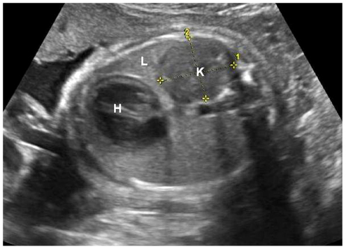
Fig. 1 Transverse sonograms of the fetal thorax on prenatal sonographic examination at 29 weeks demonstrate the ectopic kidney (K) in the left hemithorax, compressed left lung (L), mild right shift of the heart (H).
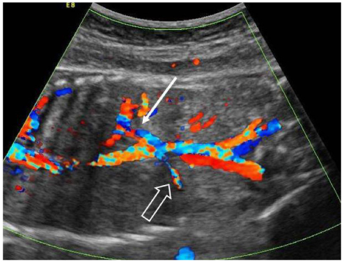
Fig. 2 Coronal sonographic view of the fetus. Color Doppler imaging demonstrates the left renal artery entering the thorax to feed the ectopic intrathoracic kidney (white arrow), and normal right renal artery entering the right kidney (hollowed arrow).
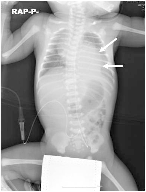
Fig. 3 Intrathoracic kidney with congenital diaphragmatic hernia on postnatal X-ray (white arrows).
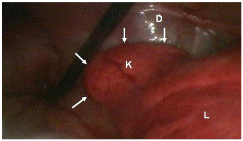
Fig. 4 Left thoracic kidney on thoracoscopy (white arrows). D, diaphragm; L, left lung; K, left kidney.
Reference
-
1. Sözübir S, Demir H, Ekingen G, Güvenç BH. Ectopic thoracic kidney in a child with congenital diaphragmatic hernia. Eur J Pediatr Surg. 2005. 15:206–209.2. Panda B, Rosenberg V, Cornfeld D, Stiller R. Prenatal diagnosis of ectopic intrathoracic kidney in a fetus with a left diaphragmatic hernia. J Clin Ultrasound. 2009. 37:47–49.3. Athanasiadis AP, Zafrakas M, Arnaoutoglou C, Karavida A, Papasozomenou P, Tarlatzis BC. Prenatal diagnosis of thoracic kidney in the 2nd trimester with delayed manifestation of associated diaphragmatic hernia. J Clin Ultrasound. 2011. 39:221–224.4. Donat SM, Donat PE. Intrathoracic kidney: a case report with a review of the world literature. J Urol. 1988. 140:131–133.5. Liddell RM, Rosenbaum DM, Blumhagen JD. Delayed radiologic appearance of bilateral thoracic ectopic kidneys. AJR Am J Roentgenol. 1989. 152:120–122.6. Yoo DG, Kim CW, Park CB, Ahn JH. Traumatic right diaphragmatic rupture combined with avulsion of the right kidney and herniation of the liver into the thorax. Korean J Thorac Cardiovasc Surg. 2011. 44:76–79.7. Hubbard AM, Crombleholme TM, Adzick NS, Coleman BG, Howell LJ, Meyer JS, et al. Prenatal MRI evaluation of congenital diaphragmatic hernia. Am J Perinatol. 1999. 16:407–413.8. Sundaram V, Vidhyashree SA, Pratap B, Surendranath A, Matthew M, Bhaskar E, et al. A male patient with right-sided thoracic kidney, diabetes mellitus, hearing loss and renal dysfunction. Int Urol Nephrol. 2007. 39:959–962.9. Singh P, Vijjan V, Gupta M, Dubey D, Srivastava A. Percutaneous nephrolithotomy of a staghorn stone in thoracic ectopic kidney. Int J Urol. 2007. 14:558–560.10. Navarro A, Jiménez J, Ríos T, Mestanza F, Aguirre I, Urquizo R. Unusual cause of lung and renal disease in a baby with trisomy 21. Pediatr Pulmonol. 2005. 40:173–174.11. Zaiss I, Kehl S, Link K, Neff W, Schaible T, Sütterlin M, et al. Associated malformations in congenital diaphragmatic hernia. Am J Perinatol. 2011. 28:211–218.12. Pelizzo G, Lembo MA, Franchella A, Giombi A, D'Agostino F, Sala S. Gastric volvulus associated with congenital diaphragmatic hernia, wandering spleen, and intrathoracic left kidney: CT findings. Abdom Imaging. 2001. 26:306–308.
- Full Text Links
-
- Actions
-
Cited
- CITED
-
- Close
- Share
-
- Similar articles
-
- Unilateral Congenital Diaphragmatic Eventration Mimicking Congenital Diaphragmatic Hernia
- Left Diaphragmatic Eventration Diagnosed as Congenital Diaphragmatic Hernia by Prenatal Sonography
- Impaction of an intrathoracic kidney acted as a shield against herniation of the abdominal viscera in a case of right congenital diaphragmatic hernia
- Congenital Thoracic Ectopic Kidney associated with Diaphragmatic Hernia in a 15-month-old Boy
- A Case of Congenital Right Diaphragmatic Eventration
